 |
PDBsum entry 1x7n

|
|
|
|
|
 |
Contents |
 |
|
|
|
|
|
|
|
|
|
|
|
|
|
|
* Residue conservation analysis
|
|
|
|
|
|
PDB id:
|
|
|
|
| Name: |
|
Isomerase
|
|
|
Title:
|
|
The crystal structure of pyrococcus furiosus phosphoglucose isomerase with bound 5-phospho-d-arabinonate and manganese
|
|
Structure:
|
|
Glucose-6-phosphate isomerase. Chain: a. Synonym: gpi, phosphoglucose isomerase, pgi, phosphohexose isomerase, phi. Engineered: yes
|
|
Source:
|
|
Pyrococcus furiosus. Organism_taxid: 2261. Gene: pgia. Expressed in: escherichia coli bl21(de3). Expression_system_taxid: 469008.
|
|
Biol. unit:
|
|
 Dimer (from PDB file)
|
|
Resolution:
|
|
|
1.89Å
|
R-factor:
|
0.144
|
R-free:
|
0.212
|
|
|
Authors:
|
|
J.M.Berrisford,J.Akerboom,S.Brouns,S.E.Sedelnikova,A.P.Turnbull,J.Van Der Oost,L.Salmon,R.Hardre,I.A.Murray,G.M.Blackburn,D.W.Rice, P.J.Baker
|
Key ref:

|
|
J.M.Berrisford
et al.
(2004).
The structures of inhibitor complexes of Pyrococcus furiosus phosphoglucose isomerase provide insights into substrate binding and catalysis.
J Mol Biol,
343,
649-657.
PubMed id:
DOI:

|
|
|
Date:
|
|
|
16-Aug-04
|
Release date:
|
12-Oct-04
|
|
|
|
|
|
|
PROCHECK
|

|
|
|
|
|
Headers
|
|
|
|
References
|
|
|
|
|
|

|
|
P83194
(G6PI_PYRFU) -
Glucose-6-phosphate isomerase from Pyrococcus furiosus (strain ATCC 43587 / DSM 3638 / JCM 8422 / Vc1)
|
|
|
|
Seq:
Struc:
|
|
|
|
189 a.a.
189 a.a.
|
|

|

|
|
|
|
|
|
|
|
|
|
|
|
Key: |
 |
PfamA domain |
|
|
 |
Secondary structure |
|
 |
CATH domain |
|
|
|
|
|
|
|
|
|
|
|
|
|
Enzyme class:
|
|
E.C.5.3.1.9
- glucose-6-phosphate isomerase.
|
|
|
|
|
|
|
Reaction:
|
|
alpha-D-glucose 6-phosphate = beta-D-fructose 6-phosphate
|
|
|
|
|
|
alpha-D-glucose 6-phosphate
Bound ligand (Het Group name = )
matches with 82.35% similarity
|
=
|
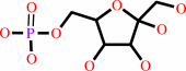 beta-D-fructose 6-phosphate
|
|
|
|
|
|
|
|
|
|
|
|
|
Molecule diagrams generated from .mol files obtained from the
KEGG ftp site
|
|
|
|
|
|
|
|
|
|
|
|
|
|
 |
|
|
|
|
|
|
| |
|
|
| |
|
DOI no:
|
J Mol Biol
343:649-657
(2004)
|
|
PubMed id:
|
|
|
|
|
|
| |
|
The structures of inhibitor complexes of Pyrococcus furiosus phosphoglucose isomerase provide insights into substrate binding and catalysis.
|
|
J.M.Berrisford,
J.Akerboom,
S.Brouns,
S.E.Sedelnikova,
A.P.Turnbull,
J.van der Oost,
L.Salmon,
R.Hardré,
I.A.Murray,
G.M.Blackburn,
D.W.Rice,
P.J.Baker.
|
|
|
|
|
| |
ABSTRACT
|
|
|
|
| |
|
|
Pyrococcus furiosus phosphoglucose isomerase (PfPGI) is a metal-containing
enzyme that catalyses the interconversion of glucose 6-phosphate (G6P) and
fructose 6-phosphate (F6P). The recent structure of PfPGI has confirmed the
hypothesis that the enzyme belongs to the cupin superfamily and identified the
position of the active site. This fold is distinct from the alphabetaalpha
sandwich fold commonly seen in phosphoglucose isomerases (PGIs) that are found
in bacteria, eukaryotes and some archaea. Whilst the mechanism of the latter
family is thought to proceed through a cis-enediol intermediate, analysis of the
structure of PfPGI in the presence of inhibitors has led to the suggestion that
the mechanism of this enzyme involves the metal-dependent direct transfer of a
hydride between C1 and C2 atoms of the substrate. To gain further insight in the
reaction mechanism of PfPGI, the structures of the free enzyme and the complexes
with the inhibitor, 5-phospho-d-arabinonate (5PAA) in the presence and absence
of metal have been determined. Comparison of these structures with those of
equivalent complexes of the eukaryotic PGIs reveals similarities at the active
site in the disposition of possible catalytic residues. These include the
presence of a glutamic acid residue, Glu97 in PfPGI, which occupies the same
position relative to the inhibitor as that of the glutamate that is thought to
function as the catalytic base in the eukaryal-type PGIs. These similarities
suggest that aspects of the catalytic mechanisms of these two structurally
unrelated PGIs may be similar and based on an enediol intermediate.
|
|
|
|
|
|
| |
Selected figure(s)
|
|
|
|
| |
|
|

|
|

|
|
Figure 1.
Figure 1. A schematic representation of the PfPGI dimer
viewed down the 2-fold axis. One subunit of the dimer is shown
in blue and red (strands and helices, respectively) and the
other in orange. The N-terminal strand can be seen to interact
with the adjacent symmetry-related strand from the other subunit
in the biological dimer.
|
|
Figure 2.
Figure 2. A stereo diagram of the active site of PfPGI
showing the 2F[O] -F[C] map, contoured at 1s (blue) and 4s
(red), electron density assigned to 5PAA in the PfPGI/5PAA
metal-free complex and its interaction with Glu97. A blue line
shows the potential hydrogen bond between the carboxyl of Glu97
and the carboxyl of 5PAA.
|
|
|
|
|
|
| |
The above figures are
reprinted
by permission from Elsevier:
J Mol Biol
(2004,
343,
649-657)
copyright 2004.
|
|
| |
Figures were
selected
by an automated process.
|
|
|
|
|
|
|
|
|
|
|
|
|
|
|
|
 |
 |
 |
|
Literature references that cite this PDB file's key reference
|
|
|
| |
PubMed id
|
|
Reference
|
|
|
|
|
|
L.M.Iyer,
S.Abhiman,
R.F.de Souza,
and
L.Aravind
(2010).
Origin and evolution of peptide-modifying dioxygenases and identification of the wybutosine hydroxylase/hydroperoxidase.
|
| |
Nucleic Acids Res,
38,
5261-5279.
|
|
|
|
|
|
|
R.K.Kuipers,
H.J.Joosten,
E.Verwiel,
S.Paans,
J.Akerboom,
J.van der Oost,
N.G.Leferink,
W.J.van Berkel,
G.Vriend,
and
P.J.Schaap
(2009).
Correlated mutation analyses on super-family alignments reveal functionally important residues.
|
| |
Proteins,
76,
608-616.
|
|
|
|
|
|
|
R.Y.Yoon,
S.J.Yeom,
C.S.Park,
and
D.K.Oh
(2009).
Substrate specificity of a glucose-6-phosphate isomerase from Pyrococcus furiosus for monosaccharides.
|
| |
Appl Microbiol Biotechnol,
83,
295-303.
|
|
|
|
|
|
|
C.Roux,
N.Gresh,
L.E.Perera,
J.P.Piquemal,
and
L.Salmon
(2007).
Binding of 5-phospho-D-arabinonohydroxamate and 5-phospho-D-arabinonate inhibitors to zinc phosphomannose isomerase from Candida albicans studied by polarizable molecular mechanics and quantum mechanics.
|
| |
J Comput Chem,
28,
938-957.
|
|
|
|
|
|
|
L.Williams,
T.Nguyen,
Y.Li,
T.N.Porter,
and
F.M.Raushel
(2006).
Uronate isomerase: a nonhydrolytic member of the amidohydrolase superfamily with an ambivalent requirement for a divalent metal ion.
|
| |
Biochemistry,
45,
7453-7462.
|
|
|
|
|
|
|
B.Siebers,
and
P.Schönheit
(2005).
Unusual pathways and enzymes of central carbohydrate metabolism in Archaea.
|
| |
Curr Opin Microbiol,
8,
695-705.
|
|
|
|
|
|
|
T.Hansen,
B.Schlichting,
J.Grötzinger,
M.K.Swan,
C.Davies,
and
P.Schönheit
(2005).
Mutagenesis of catalytically important residues of cupin type phosphoglucose isomerase from Archaeoglobus fulgidus.
|
| |
FEBS J,
272,
6266-6275.
|
|
|
|
|
|
|
T.Hansen,
and
P.Schönheit
(2005).
Escherichia coli phosphoglucose isomerase can be substituted by members of the PGI family, the PGI/PMI family, and the cPGI family.
|
| |
FEMS Microbiol Lett,
250,
49-53.
|
|
|
|
|
|
The most recent references are shown first.
Citation data come partly from CiteXplore and partly
from an automated harvesting procedure. Note that this is likely to be
only a partial list as not all journals are covered by
either method. However, we are continually building up the citation data
so more and more references will be included with time.
|
|


 Links
Links


