 |
PDBsum entry 1vq3

|
|
|
|
|
 |
Contents |
 |
|
|
|
|
|
|
|
|
|
|
* Residue conservation analysis
|
|
|
|
|
|
|
|
|
|
|
Enzyme class:
|
|
E.C.6.3.5.3
- phosphoribosylformylglycinamidine synthase.
|
|
|
|
|
|
|
Pathway:
|
|
Purine Biosynthesis (early stages)
|
|
|
|
|
|
Reaction:
|
|
N2-formyl-N1-(5-phospho-beta-D-ribosyl)glycinamide + L-glutamine + ATP + H2O = 2-formamido-N1-(5-O-phospho-beta-D-ribosyl)acetamidine + L-glutamate + ADP + phosphate + H+
|
|
|
|
|
|
N(2)-formyl-N(1)-(5-phospho-beta-D-ribosyl)glycinamide
|
+
|
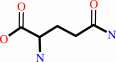 L-glutamine
|
+
|
 ATP
ATP
|
+
|
H2O
|
=
|
2-formamido-N(1)-(5-O-phospho-beta-D-ribosyl)acetamidine
|
+
|
 L-glutamate
|
+
|
 ADP
ADP
|
+
|
 phosphate
|
+
|
H(+)
|
|
|
|
|
|
|
|
|
|
|
|
|
Molecule diagrams generated from .mol files obtained from the
KEGG ftp site
|
|
|
|
|
|
|
|
|
|
|
|
|
|
 |
|
|
|
|
|
|
| |
|
|
| |
|
DOI no:
|
Proteins
65:249-254
(2006)
|
|
PubMed id:
|
|
|
|
|
|
| |
|
Crystal structure of phosphoribosylformyl-glycinamidine synthase II, PurS subunit (TM1244) from Thermotoga maritima at 1.90 A resolution.
|
|
I.I.Mathews,
S.S.Krishna,
R.Schwarzenbacher,
D.McMullan,
L.Jaroszewski,
M.D.Miller,
P.Abdubek,
S.Agarwalla,
E.Ambing,
H.L.Axelrod,
J.M.Canaves,
D.Carlton,
H.J.Chiu,
T.Clayton,
M.DiDonato,
L.Duan,
M.A.Elsliger,
S.K.Grzechnik,
J.Hale,
E.Hampton,
J.Haugen,
K.K.Jin,
H.E.Klock,
E.Koesema,
J.S.Kovarik,
A.Kreusch,
P.Kuhn,
I.Levin,
A.T.Morse,
E.Nigoghossian,
L.Okach,
S.Oommachen,
J.Paulsen,
K.Quijano,
R.Reyes,
C.L.Rife,
G.Spraggon,
R.C.Stevens,
H.van den Bedem,
A.White,
G.Wolf,
Q.Xu,
K.O.Hodgson,
J.Wooley,
A.M.Deacon,
A.Godzik,
S.A.Lesley,
I.A.Wilson.
|
|
|
|
|
| |
ABSTRACT
|
|
|
|
| |
|
|
|
|
| |
Selected figure(s)
|
|
|
|
| |
|
|

|
|

|
|
Figure 2.
Figure 2. Domain delineation of PurS. (A) Structural
representative of the ferredoxin-like fold: the
anticodon-binding domain of PheRS (PDB code: 1pys, chain B). (B)
Dimeric unit of PurS colored according to chains (chain A: blue,
chain B: red). The PurS domain-swapped dimer structure contains
two domains (separated by a black horizontal bar), each of which
resembles the ferredoxin-like fold. The ferredoxin-like fold is
made of four  -strands
and two -strands
and two  -helices,
and in PurS these structural elements are contributed from the
two chains of the dimer by a domain swap of -helix
H1 and -strand
2.
The elements are labeled according to the ferredoxin-like fold. -helices,
and in PurS these structural elements are contributed from the
two chains of the dimer by a domain swap of -helix
H1 and -strand
2.
The elements are labeled according to the ferredoxin-like fold.
|
|
Figure 3.
Figure 3. (A) Structural alignment of PurS (blue) with the
N-terminal domain of lgPurL (gray) from S. typhimurium C[  ]atoms
from the dimer of TM1244 were used for the alignment with
lgPurL. The largest structural difference corresponds to strand
3
of chain A, which is replaced by a long loop (see arrow) that
acts as a linker between the adjacent PurS-like domains in
lgPurL. (B) Stereo ribbon diagram of a superposition of lgPurL
from S. typhimurium (FGAR-AT; gray) with PurS (TM1244; blue) and
smPurL (TM1246; orange). Ligands of lgPurL are shown as CPK
(Corey, Pauling, and Kultun) color scheme. The arrow indicates
the expected location of the PurQ protein (TM1245) in FGAR-AT
from T. maritima. ]atoms
from the dimer of TM1244 were used for the alignment with
lgPurL. The largest structural difference corresponds to strand
3
of chain A, which is replaced by a long loop (see arrow) that
acts as a linker between the adjacent PurS-like domains in
lgPurL. (B) Stereo ribbon diagram of a superposition of lgPurL
from S. typhimurium (FGAR-AT; gray) with PurS (TM1244; blue) and
smPurL (TM1246; orange). Ligands of lgPurL are shown as CPK
(Corey, Pauling, and Kultun) color scheme. The arrow indicates
the expected location of the PurQ protein (TM1245) in FGAR-AT
from T. maritima.
|
|
|
|
|
|
| |
The above figures are
reprinted
by permission from John Wiley & Sons, Inc.:
Proteins
(2006,
65,
249-254)
copyright 2006.
|
|
| |
Figures were
selected
by an automated process.
|
|
|
|
|
|
|
|
|
|
|
|
|
|
|
|
 |
 |
 |
|
Literature references that cite this PDB file's key reference
|
|
|
| |
PubMed id
|
|
Reference
|
|
|
|
|
|
H.L.Axelrod,
D.McMullan,
S.S.Krishna,
M.D.Miller,
M.A.Elsliger,
P.Abdubek,
E.Ambing,
T.Astakhova,
D.Carlton,
H.J.Chiu,
T.Clayton,
L.Duan,
J.Feuerhelm,
S.K.Grzechnik,
J.Hale,
G.W.Han,
J.Haugen,
L.Jaroszewski,
K.K.Jin,
H.E.Klock,
M.W.Knuth,
E.Koesema,
A.T.Morse,
E.Nigoghossian,
L.Okach,
S.Oommachen,
J.Paulsen,
K.Quijano,
R.Reyes,
C.L.Rife,
H.van den Bedem,
D.Weekes,
A.White,
G.Wolf,
Q.Xu,
K.O.Hodgson,
J.Wooley,
A.M.Deacon,
A.Godzik,
S.A.Lesley,
and
I.A.Wilson
(2008).
Crystal structure of AICAR transformylase IMP cyclohydrolase (TM1249) from Thermotoga maritima at 1.88 A resolution.
|
| |
Proteins,
71,
1042-1049.
|
|
|
PDB code:
|
|
|
|
|
|
|
|
M.Morar,
A.A.Hoskins,
J.Stubbe,
and
S.E.Ealick
(2008).
Formylglycinamide ribonucleotide amidotransferase from Thermotoga maritima: structural insights into complex formation.
|
| |
Biochemistry,
47,
7816-7830.
|
|
|
PDB code:
|
|
|
|
|
|
|
|
Y.Zhang,
M.Morar,
and
S.E.Ealick
(2008).
Structural biology of the purine biosynthetic pathway.
|
| |
Cell Mol Life Sci,
65,
3699-3724.
|
|
|
|
|
|
The most recent references are shown first.
Citation data come partly from CiteXplore and partly
from an automated harvesting procedure. Note that this is likely to be
only a partial list as not all journals are covered by
either method. However, we are continually building up the citation data
so more and more references will be included with time.
Where a reference describes a PDB structure, the PDB
code is
shown on the right.
|
|


 Links
Links


