 |
PDBsum entry 1vbg

|
|
|
|
|
 |
Contents |
 |
|
|
|
|
|
|
|
|
|
|
|
|
|
|
* Residue conservation analysis
|
|
|
|
|
|
PDB id:
|
|
|
|
| Name: |
|
Transferase
|
|
|
Title:
|
|
Pyruvate phosphate dikinase from maize
|
|
Structure:
|
|
Pyruvate,orthophosphate dikinase. Chain: a. Synonym: pyruvate phosphate dikinase. Engineered: yes
|
|
Source:
|
|
Zea mays. Organism_taxid: 4577. Expressed in: escherichia coli. Expression_system_taxid: 562.
|
|
Biol. unit:
|
|
 Dimer (from PDB file)
|
|
Resolution:
|
|
|
2.30Å
|
R-factor:
|
0.209
|
R-free:
|
0.237
|
|
|
Authors:
|
|
T.Nakanishi,T.Nakatsu,M.Matsuoka,K.Sakata,H.Kato,Riken Structural Genomics/proteomics Initiative (Rsgi)
|
Key ref:

|
|
T.Nakanishi
et al.
(2005).
Crystal structures of pyruvate phosphate dikinase from maize revealed an alternative conformation in the swiveling-domain motion.
Biochemistry,
44,
1136-1144.
PubMed id:
DOI:
|
|
|
Date:
|
|
|
26-Feb-04
|
Release date:
|
08-Mar-05
|
|
|
|
|
|
|
PROCHECK
|

|
|
|
|
|
Headers
|
|
|
|
References
|
|
|
|
|
|

|
|
P11155
(PPDK1_MAIZE) -
Pyruvate, phosphate dikinase 1, chloroplastic from Zea mays
|
|
|
|
Seq:
Struc:
|
|
|
|
947 a.a.
874 a.a.*
|
|

|

|

|
|
|
|
|
|
|
|
|
|
|
Key: |
 |
PfamA domain |
|
|
 |
Secondary structure |
|
 |
CATH domain |
|
|
*
PDB and UniProt seqs differ
at 8 residue positions (black
crosses)
|
|
|
|
|
|
|
|
|
|
|
|
|
Enzyme class:
|
|
E.C.2.7.9.1
- pyruvate, phosphate dikinase.
|
|
|
|
|
|
|
Reaction:
|
|
pyruvate + phosphate + ATP = phosphoenolpyruvate + AMP + diphosphate + H+
|
|
|
|
|
|
 pyruvate
|
+
|
 phosphate
|
+
|
 ATP
ATP
|
=
|
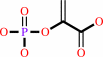 phosphoenolpyruvate
|
+
|
 AMP
AMP
|
+
|
 diphosphate
|
+
|
H(+)
|
|
|
|
|
|
|
|
|
|
|
|
|
Molecule diagrams generated from .mol files obtained from the
KEGG ftp site
|
|
|
|
|
|
|
|
|
|
|
|
|
|
 |
|
|
|
|
|
|
| |
|
|
| |
|
DOI no:
|
Biochemistry
44:1136-1144
(2005)
|
|
PubMed id:
|
|
|
|
|
|
| |
|
Crystal structures of pyruvate phosphate dikinase from maize revealed an alternative conformation in the swiveling-domain motion.
|
|
T.Nakanishi,
T.Nakatsu,
M.Matsuoka,
K.Sakata,
H.Kato.
|
|
|
|
|
| |
ABSTRACT
|
|
|
|
| |
|
|
Pyruvate phosphate dikinase (PPDK) reversibly catalyzes the conversion of ATP,
phosphate, and pyruvate into AMP, pyrophosphate, and phosphoenolpyruvate (PEP),
respectively. Since the nucleotide binding site (in the N-terminal domain) and
the pyruvate/PEP binding site (in the C-terminal domain) are separated by
approximately 45 A, it has been proposed that an intermediary domain, called the
central domain, swivels between these remote domains to transfer the phosphate.
However, no direct structural evidence for the swiveling central domain has been
found. In this study, the crystal structures of maize PPDK with and without PEP
have been determined at 2.3 A resolution. These structures revealed that the
central domain is located near the pyruvate/PEP binding C-terminal domain, in
contrast to the PPDK from Clostridium symbiosum, wherein the central domain is
located near the nucleotide-binding N-terminal domain. Structural comparisons
between the maize and C. symbiosum PPDKs demonstrated that the swiveling motion
of the central domain consists of a rotation of at least 92 degrees and a
translation of 0.5 A. By comparing the maize PPDK structures with and without
PEP, we have elucidated the mode of binding of PEP to the C-terminal domain and
the induced conformational changes in the central domain.
|
|
|
|
|
|
|
|
|
|
|
|
|
|
|
|
|
|
 |
 |
 |
|
Literature references that cite this PDB file's key reference
|
|
|
| |
PubMed id
|
|
Reference
|
|
|
|
|
|
A.Teplyakov,
K.Lim,
P.P.Zhu,
G.Kapadia,
C.C.Chen,
J.Schwartz,
A.Howard,
P.T.Reddy,
A.Peterkofsky,
and
O.Herzberg
(2006).
Structure of phosphorylated enzyme I, the phosphoenolpyruvate:sugar phosphotransferase system sugar translocation signal protein.
|
| |
Proc Natl Acad Sci U S A,
103,
16218-16223.
|
|
|
PDB code:
|
|
|
|
|
|
|
|
E.Hurtado-Gómez,
G.Fernández-Ballester,
H.Nothaft,
J.Gómez,
F.Titgemeyer,
and
J.L.Neira
(2006).
Biophysical characterization of the enzyme I of the Streptomyces coelicolor phosphoenolpyruvate:sugar phosphotransferase system.
|
| |
Biophys J,
90,
4592-4604.
|
|
|
|
|
|
|
J.Deutscher,
C.Francke,
and
P.W.Postma
(2006).
How phosphotransferase system-related protein phosphorylation regulates carbohydrate metabolism in bacteria.
|
| |
Microbiol Mol Biol Rev,
70,
939.
|
|
|
|
|
|
|
J.Márquez,
S.Reinelt,
B.Koch,
R.Engelmann,
W.Hengstenberg,
and
K.Scheffzek
(2006).
Structure of the full-length enzyme I of the phosphoenolpyruvate-dependent sugar phosphotransferase system.
|
| |
J Biol Chem,
281,
32508-32515.
|
|
|
PDB code:
|
|
|
|
|
|
|
The most recent references are shown first.
Citation data come partly from CiteXplore and partly
from an automated harvesting procedure. Note that this is likely to be
only a partial list as not all journals are covered by
either method. However, we are continually building up the citation data
so more and more references will be included with time.
Where a reference describes a PDB structure, the PDB
code is
shown on the right.
|
|


 Links
Links


