 |
PDBsum entry 1rk4

|
|
|
|
|
|

|

|

|

|
|

|
|

|
|

|
|

|
|

|
|
|
 |

|

|
|
|
|
|
|
|
|
|
|
|
|
|
|
|
|
|
|
|
|
|
|
|
|
|
|
|
|
|
|
|
|
|
|
Ion transport/membrane protein
|
PDB id
|
|
|
|
1rk4
|
|
|
|
|
|
|
|
|
|
|
|
|
|
|

|

|

|
|
|
|
|
|
|
|
 |
Contents |
 |
|
|
|
|
|
|
|
|
|
|
* Residue conservation analysis
|
|
|
|
|
|
PDB id:
|
|
|
|
| Name: |
|
Ion transport/membrane protein
|
|
|
Title:
|
|
Crystal structure of a soluble dimeric form of oxidised clic1
|
|
Structure:
|
|
Chloride intracellular channel protein 1. Chain: a, b. Synonym: nuclear chloride ion channel 27. Ncc27. P64 clcp. Chloride channel abp. P64clcp. Chloride channel abp. Engineered: yes
|
|
Source:
|
|
Homo sapiens. Human. Organism_taxid: 9606. Gene: clic1, ncc27. Expressed in: escherichia coli. Expression_system_taxid: 562.
|
|
Biol. unit:
|
|
Dimer (from
)
|
|
Resolution:
|
|
|
1.79Å
|
R-factor:
|
0.178
|
R-free:
|
0.213
|
|
|
Authors:
|
|
D.R.Littler,S.J.Harrop,W.D.Fairlie,L.J.Brown,G.J.Pankhurst, S.Pankhurst,M.Z.Demaere,T.J.Campbell,A.R.Bauskin,R.Tonini, M.Mazzanti,S.N.Breit,P.M.Curmi
|
Key ref:

|
|
D.R.Littler
et al.
(2004).
The intracellular chloride ion channel protein CLIC1 undergoes a redox-controlled structural transition.
J Biol Chem,
279,
9298-9305.
PubMed id:
DOI:

|
|
|
Date:
|
|
|
20-Nov-03
|
Release date:
|
02-Dec-03
|
|
|
|
|
|
|
PROCHECK
|

|
|
|
|
|
Headers
|
|
|
|
References
|
|
|
|
|
|

|
|
O00299
(CLIC1_HUMAN) -
Chloride intracellular channel protein 1 from Homo sapiens
|
|
|
|
Seq:
Struc:
|
|
|
|
241 a.a.
213 a.a.
|
|

|

|
|
|
|
|
|
|
|
|
|
|
|
Key: |
 |
PfamA domain |
|
|
 |
Secondary structure |
|
 |
CATH domain |
|
|
|
|
|
|
|
|
|
|
|
|
|
Enzyme class 2:
|
|
E.C.1.8.-.-
|
|
|
|
|
|
|
Enzyme class 3:
|
|
E.C.1.8.5.1
- glutathione dehydrogenase (ascorbate).
|
|
|
|
|
|
|
Reaction:
|
|
L-dehydroascorbate + 2 glutathione = glutathione disulfide + L-ascorbate
|
|
|
|
|
|
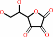 L-dehydroascorbate
|
+
|
 2
×
glutathione
2
×
glutathione
|
=
|
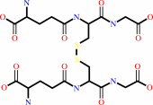 glutathione disulfide
|
+
|
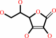 L-ascorbate
|
|
|
|
|
|
|
|
|
|
|
|
|
Note, where more than one E.C. class is given (as above), each may
correspond to a different protein domain or, in the case of polyprotein
precursors, to a different mature protein.
|
|
|
|
Molecule diagrams generated from .mol files obtained from the
KEGG ftp site
|
|
|
|
|
|
|
|
|
|
|
|
|
|
 |
|
|
|
|
|
|
| |
|
|
| |
|
DOI no:
|
J Biol Chem
279:9298-9305
(2004)
|
|
PubMed id:
|
|
|
|
|
|
| |
|
The intracellular chloride ion channel protein CLIC1 undergoes a redox-controlled structural transition.
|
|
D.R.Littler,
S.J.Harrop,
W.D.Fairlie,
L.J.Brown,
G.J.Pankhurst,
S.Pankhurst,
M.Z.DeMaere,
T.J.Campbell,
A.R.Bauskin,
R.Tonini,
M.Mazzanti,
S.N.Breit,
P.M.Curmi.
|
|
|
|
|
| |
ABSTRACT
|
|
|
|
| |
|
|
Most proteins adopt a well defined three-dimensional structure; however, it is
increasingly recognized that some proteins can exist with at least two stable
conformations. Recently, a class of intracellular chloride ion channel proteins
(CLICs) has been shown to exist in both soluble and integral membrane forms. The
structure of the soluble form of CLIC1 is typical of a soluble glutathione
S-transferase superfamily protein but contains a glutaredoxin-like active site.
In this study we show that on oxidation CLIC1 undergoes a reversible transition
from a monomeric to a non-covalent dimeric state due to the formation of an
intramolecular disulfide bond (Cys-24-Cys-59). We have determined the crystal
structure of this oxidized state and show that a major structural transition has
occurred, exposing a large hydrophobic surface, which forms the dimer interface.
The oxidized CLIC1 dimer maintains its ability to form chloride ion channels in
artificial bilayers and vesicles, whereas a reducing environment prevents the
formation of ion channels by CLIC1. Mutational studies show that both Cys-24 and
Cys-59 are required for channel activity.
|
|
|
|
|
|
| |
Selected figure(s)
|
|
|
|
| |
|
|

|
|

|
|
Figure 2.
FIG. 2. Structure of the oxidized CLIC1 dimer. A, stereo
backbone of the CLIC1 dimer; green, A subunit; red, B subunit.
In the A subunit every 10th residue is labeled. B, electron
density of the intramolecular disulfide bond between Cys-24 and
Cys-59 contoured at 1  . The CLIC1 dimer is
viewed along (C) and perpendicular (D) to the pseudo 2-fold
axis; are shown helices, A subunit (red) and B subunit (green),
and intramolecular disulfide bonds (yellow). E, ClustalW (24)
alignment of the CLIC family. The secondary structure is shown
for both monomeric (red, helices; yellow, . The CLIC1 dimer is
viewed along (C) and perpendicular (D) to the pseudo 2-fold
axis; are shown helices, A subunit (red) and B subunit (green),
and intramolecular disulfide bonds (yellow). E, ClustalW (24)
alignment of the CLIC family. The secondary structure is shown
for both monomeric (red, helices; yellow,  -strands) and dimeric
(blue, helices) forms. Conserved regions are shaded; green,
putative transmembrane regions; yellow, Cys; cream, Gly.
Features unique to CLIC1 are in blue. Ramachandran distances
(see "Experimental Procedures") for the monomer to dimer
transition are plotted above its sequence. Figures were made
with SETOR (25), MOLSCRIPT (26), RASTER3D (27), and CONSCRIPT
(28). -strands) and dimeric
(blue, helices) forms. Conserved regions are shaded; green,
putative transmembrane regions; yellow, Cys; cream, Gly.
Features unique to CLIC1 are in blue. Ramachandran distances
(see "Experimental Procedures") for the monomer to dimer
transition are plotted above its sequence. Figures were made
with SETOR (25), MOLSCRIPT (26), RASTER3D (27), and CONSCRIPT
(28).
|
|
Figure 3.
FIG. 3. Structural transition of CLIC1 between the
monomeric and the dimeric forms. Representations of reduced
monomeric form of CLIC1 (A) and a subunit of the oxidized
dimeric form (B). C, backbone superposition of CLIC1 for the
reduced monomeric (green) and the oxidized dimeric (magenta)
states. Ramachandran distances for residues 23-234 are mapped
onto the backbone of the monomeric (D) and dimeric (E) forms.
The color gradient, from gray to pink represents Ramachandran
distances from 0° to 180°. Residues not observed in the
dimer are colored gold. F, Ramachandran plot of residues within
the N-domain with Ramachandran distances greater than 35^0
between the two structures. Monomer  - -  co-ordinates are plotted
as orange squares, and dimer co-ordinates are in black with a
connecting line. The figures were made with SETOR (25),
MOLSCRIPT (26), RASTER3D (27), and GRASP (29). co-ordinates are plotted
as orange squares, and dimer co-ordinates are in black with a
connecting line. The figures were made with SETOR (25),
MOLSCRIPT (26), RASTER3D (27), and GRASP (29).
|
|
|
|
|
|
| |
The above figures are
reprinted
by permission from the ASBMB:
J Biol Chem
(2004,
279,
9298-9305)
copyright 2004.
|
|
| |
Figures were
selected
by the author.
|
|
|
|
|
|
|
|
|
|
|
|
|
|
|
|
 |
 |
 |
|
Literature references that cite this PDB file's key reference
|
|
|
| |
PubMed id
|
|
Reference
|
|
|
|
|
|
A.F.Dulhunty,
R.Hewawasam,
D.Liu,
M.G.Casarotto,
and
P.G.Board
(2011).
Regulation of the cardiac muscle ryanodine receptor by glutathione transferases.
|
| |
Drug Metab Rev,
43,
236-252.
|
|
|
|
|
|
|
F.Sesti,
S.Liu,
and
S.Q.Cai
(2010).
Oxidation of potassium channels by ROS: a general mechanism of aging and neurodegeneration?
|
| |
Trends Cell Biol,
20,
45-51.
|
|
|
|
|
|
|
J.J.Tung,
and
J.Kitajewski
(2010).
Chloride intracellular channel 1 functions in endothelial cell growth and migration.
|
| |
J Angiogenes Res,
2,
23.
|
|
|
|
|
|
|
M.A.Wouters,
S.W.Fan,
and
N.L.Haworth
(2010).
Disulfides as redox switches: from molecular mechanisms to functional significance.
|
| |
Antioxid Redox Signal,
12,
53-91.
|
|
|
|
|
|
|
P.N.Bryan,
and
J.Orban
(2010).
Proteins that switch folds.
|
| |
Curr Opin Struct Biol,
20,
482-488.
|
|
|
|
|
|
|
R.Brauer,
L.C.Wang,
S.T.Woon,
D.J.Bridewell,
K.Henare,
D.Malinger,
B.D.Palmer,
S.N.Vogel,
C.Kieda,
S.M.Tijono,
and
L.M.Ching
(2010).
Labeling of oxidizable proteins with a photoactivatable analog of the antitumor agent DMXAA: evidence for redox signaling in its mode of action.
|
| |
Neoplasia,
12,
755-765.
|
|
|
|
|
|
|
A.Shukla,
M.Malik,
C.Cataisson,
Y.Ho,
T.Friesen,
K.S.Suh,
and
S.H.Yuspa
(2009).
TGF-beta signalling is regulated by Schnurri-2-dependent nuclear translocation of CLIC4 and consequent stabilization of phospho-Smad2 and 3.
|
| |
Nat Cell Biol,
11,
777-784.
|
|
|
|
|
|
|
B.Ponsioen,
L.van Zeijl,
M.Langeslag,
M.Berryman,
D.Littler,
K.Jalink,
and
W.H.Moolenaar
(2009).
Spatiotemporal regulation of chloride intracellular channel protein CLIC4 by RhoA.
|
| |
Mol Biol Cell,
20,
4664-4672.
|
|
|
|
|
|
|
S.H.Stoychev,
C.Nathaniel,
S.Fanucchi,
M.Brock,
S.Li,
K.Asmus,
V.L.Woods,
and
H.W.Dirr
(2009).
Structural dynamics of soluble chloride intracellular channel protein CLIC1 examined by amide hydrogen-deuterium exchange mass spectrometry.
|
| |
Biochemistry,
48,
8413-8421.
|
|
|
|
|
|
|
S.W.Fan,
R.A.George,
N.L.Haworth,
L.L.Feng,
J.Y.Liu,
and
M.A.Wouters
(2009).
Conformational changes in redox pairs of protein structures.
|
| |
Protein Sci,
18,
1745-1765.
|
|
|
|
|
|
|
X.Meng,
G.Wang,
C.Viero,
Q.Wang,
W.Mi,
X.D.Su,
T.Wagenknecht,
A.J.Williams,
Z.Liu,
and
C.C.Yin
(2009).
CLIC2-RyR1 interaction and structural characterization by cryo-electron microscopy.
|
| |
J Mol Biol,
387,
320-334.
|
|
|
|
|
|
|
A.Rahman,
S.G.Kumar,
S.W.Kim,
H.J.Hwang,
Y.M.Baek,
S.H.Lee,
H.S.Hwang,
Y.H.Shon,
K.S.Nam,
and
J.W.Yun
(2008).
Proteomic analysis for inhibitory effect of chitosan oligosaccharides on 3T3-L1 adipocyte differentiation.
|
| |
Proteomics,
8,
569-581.
|
|
|
|
|
|
|
D.R.Littler,
S.J.Harrop,
L.J.Brown,
G.J.Pankhurst,
A.V.Mynott,
P.Luciani,
R.A.Mandyam,
M.Mazzanti,
S.Tanda,
M.A.Berryman,
S.N.Breit,
and
P.M.Curmi
(2008).
Comparison of vertebrate and invertebrate CLIC proteins: the crystal structures of Caenorhabditis elegans EXC-4 and Drosophila melanogaster DmCLIC.
|
| |
Proteins,
71,
364-378.
|
|
|
PDB codes:
|
|
|
|
|
|
|
|
R.R.Thangudu,
M.Manoharan,
N.Srinivasan,
F.Cadet,
R.Sowdhamini,
and
B.Offmann
(2008).
Analysis on conservation of disulphide bonds and their structural features in homologous protein domain families.
|
| |
BMC Struct Biol,
8,
55.
|
|
|
|
|
|
|
B.A.Cromer,
M.A.Gorman,
G.Hansen,
J.J.Adams,
M.Coggan,
P.G.Board,
and
M.W.Parker
(2007).
Expression, purification, crystallization and preliminary X-ray diffraction analysis of chloride intracellular channel 2 (CLIC2).
|
| |
Acta Crystallogr Sect F Struct Biol Cryst Commun,
63,
961-963.
|
|
|
|
|
|
|
B.Ulmasov,
J.Bruno,
P.G.Woost,
and
J.C.Edwards
(2007).
Tissue and subcellular distribution of CLIC1.
|
| |
BMC Cell Biol,
8,
8.
|
|
|
|
|
|
|
C.S.Sevier,
and
C.A.Kaiser
(2006).
Disulfide transfer between two conserved cysteine pairs imparts selectivity to protein oxidation by Ero1.
|
| |
Mol Biol Cell,
17,
2256-2266.
|
|
|
|
|
|
|
H.Singh,
and
R.H.Ashley
(2006).
Redox regulation of CLIC1 by cysteine residues associated with the putative channel pore.
|
| |
Biophys J,
90,
1628-1638.
|
|
|
|
|
|
|
J.C.Edwards
(2006).
The CLIC1 chloride channel is regulated by the cystic fibrosis transmembrane conductance regulator when expressed in Xenopus oocytes.
|
| |
J Membr Biol,
213,
39-46.
|
|
|
|
|
|
|
D.R.Littler,
N.N.Assaad,
S.J.Harrop,
L.J.Brown,
G.J.Pankhurst,
P.Luciani,
M.I.Aguilar,
M.Mazzanti,
M.A.Berryman,
S.N.Breit,
and
P.M.Curmi
(2005).
Crystal structure of the soluble form of the redox-regulated chloride ion channel protein CLIC4.
|
| |
FEBS J,
272,
4996-5007.
|
|
|
PDB code:
|
|
|
|
|
|
|
|
J.D.Hayes,
J.U.Flanagan,
and
I.R.Jowsey
(2005).
Glutathione transferases.
|
| |
Annu Rev Pharmacol Toxicol,
45,
51-88.
|
|
|
|
|
|
|
N.J.Marianayagam,
M.Sunde,
and
J.M.Matthews
(2004).
The power of two: protein dimerization in biology.
|
| |
Trends Biochem Sci,
29,
618-625.
|
|
|
|
|
|
The most recent references are shown first.
Citation data come partly from CiteXplore and partly
from an automated harvesting procedure. Note that this is likely to be
only a partial list as not all journals are covered by
either method. However, we are continually building up the citation data
so more and more references will be included with time.
Where a reference describes a PDB structure, the PDB
codes are
shown on the right.
|
|


 Links
Links


