 |
PDBsum entry 1rf6

|
|
|
|
|
 |
Contents |
 |
|
|
|
|
|
|
|
|
|
|
|
|
* Residue conservation analysis
|
|
|
|
|
|
PDB id:
|
|
|
|
| Name: |
|
Transferase
|
|
|
Title:
|
|
Structural studies of streptococcus pneumoniae epsp synthase in s3p- glp bound state
|
|
Structure:
|
|
5-enolpyruvylshikimate-3-phosphate synthase. Chain: a, b, c, d. Engineered: yes
|
|
Source:
|
|
Streptococcus pneumoniae. Organism_taxid: 1313. Gene: aroa. Expressed in: escherichia coli. Expression_system_taxid: 562
|
|
Resolution:
|
|
|
1.90Å
|
R-factor:
|
0.218
|
R-free:
|
0.229
|
|
|
Authors:
|
|
H.Park,J.L.Hilsenbeck,H.J.Kim,W.A.Shuttleworth,Y.H.Park,J.N.Evans, C.Kang
|
|
Key ref:
|
|
H.Park
et al.
(2004).
Structural studies of Streptococcus pneumoniae EPSP synthase in unliganded state, tetrahedral intermediate-bound state and S3P-GLP-bound state.
Mol Microbiol,
51,
963-971.
PubMed id:
|
|
|
Date:
|
|
|
07-Nov-03
|
Release date:
|
17-Feb-04
|
|
|
|
|
|
|
PROCHECK
|

|
|
|
|
|
Headers
|
|
|
|
References
|
|
|
|
|
|

|
|
Q9S400
(AROA_STRPN) -
3-phosphoshikimate 1-carboxyvinyltransferase from Streptococcus pneumoniae serotype 4 (strain ATCC BAA-334 / TIGR4)
|
|
|
|
Seq:
Struc:
|
|
|
|
427 a.a.
427 a.a.*
|
|

|

|

|
|
|
|
|
|
|
|
|
|
|
Key: |
 |
PfamA domain |
|
|
 |
Secondary structure |
|
 |
CATH domain |
|
|
*
PDB and UniProt seqs differ
at 4 residue positions (black
crosses)
|
|
|
|
|
|
|
|
|
|
|
|
|
Enzyme class:
|
|
E.C.2.5.1.19
- 3-phosphoshikimate 1-carboxyvinyltransferase.
|
|
|
|
|
|
|
Pathway:
|
|
Shikimate and Chorismate Biosynthesis
|
|
|
|
|
|
Reaction:
|
|
3-phosphoshikimate + phosphoenolpyruvate = 5-O-(1-carboxyvinyl)-3- phosphoshikimate + phosphate
|
|
|
|
|
|
3-phosphoshikimate
|
+
|
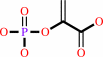 phosphoenolpyruvate
|
=
|
5-O-(1-carboxyvinyl)-3- phosphoshikimate
Bound ligand (Het Group name = )
matches with 76.19% similarity
|
+
|
 phosphate
|
|
|
|
|
|
|
|
|
|
|
|
|
Molecule diagrams generated from .mol files obtained from the
KEGG ftp site
|
|
|
|
|
|
|
|
|
|
|
|
|
|
 |
|
|
|
|
|
|
| |
|
|
| |
|
|
Mol Microbiol
51:963-971
(2004)
|
|
PubMed id:
|
|
|
|
|
|
| |
|
Structural studies of Streptococcus pneumoniae EPSP synthase in unliganded state, tetrahedral intermediate-bound state and S3P-GLP-bound state.
|
|
H.Park,
J.L.Hilsenbeck,
H.J.Kim,
W.A.Shuttleworth,
Y.H.Park,
J.N.Evans,
C.Kang.
|
|
|
|
|
| |
ABSTRACT
|
|
|
|
| |
|
|
The shikimate pathway synthesizes aromatic amino acids and other essential
metabolites that are necessary for bacteria, plants and fungi to survive. This
pathway is not present in vertebrates and therefore represents an attractive
target for antibacterial agents. We have successfully crystallized and solved
the structure of unliganded, inhibitor-liganded and tetrahedral intermediate
(TI)-liganded forms of Streptococcus pneumoniae EPSP synthase. The overall
topology of the S. pneumoniae EPSP synthase is similar to that of the
Escherichia coli EPSP synthase. In addition, the majority of residues
responsible for ligand binding were conserved between the two proteins.
TI-liganded structure provides absolute configuration of the C-2 atom from the
F-PEP moiety of the enzyme-bound intermediate and also defines key residues
responsible for the enzyme reaction. Comparison of the unliganded state and
substrate-bound state of the enzyme provides insights into the structural
mechanisms involved in dynamic events of ligand binding, domain movement and
closure. This structural study of the pathogenic bacteria S. pneumoniae EPSP
synthase with inhibitor and TI will provide invaluable information for the
design of new-generation antibiotics.
|
|
|
|
|
|
|
|
|
|
|
|
|
|
|
|
|
|
 |
 |
 |
|
Literature references that cite this PDB file's key reference
|
|
|
| |
PubMed id
|
|
Reference
|
|
|
|
|
|
T.Funke,
Y.Yang,
H.Han,
M.Healy-Fried,
S.Olesen,
A.Becker,
and
E.Schönbrunn
(2009).
Structural basis of glyphosate resistance resulting from the double mutation Thr97 -> Ile and Pro101 -> Ser in 5-enolpyruvylshikimate-3-phosphate synthase from Escherichia coli.
|
| |
J Biol Chem,
284,
9854-9860.
|
|
|
PDB codes:
|
|
|
|
|
|
|
|
M.Brylinski,
and
J.Skolnick
(2008).
What is the relationship between the global structures of apo and holo proteins?
|
| |
Proteins,
70,
363-377.
|
|
|
|
|
|
|
S.O.Yesylevskyy,
V.N.Kharkyanen,
and
A.P.Demchenko
(2008).
The blind search for the closed states of hinge-bending proteins.
|
| |
Proteins,
71,
831-843.
|
|
|
|
|
|
|
M.L.Healy-Fried,
T.Funke,
M.A.Priestman,
H.Han,
and
E.Schönbrunn
(2007).
Structural basis of glyphosate tolerance resulting from mutations of Pro101 in Escherichia coli 5-enolpyruvylshikimate-3-phosphate synthase.
|
| |
J Biol Chem,
282,
32949-32955.
|
|
|
PDB codes:
|
|
|
|
|
|
|
|
T.Funke,
H.Han,
M.L.Healy-Fried,
M.Fischer,
and
E.Schönbrunn
(2006).
Molecular basis for the herbicide resistance of Roundup Ready crops.
|
| |
Proc Natl Acad Sci U S A,
103,
13010-13015.
|
|
|
PDB codes:
|
|
|
|
|
|
|
|
Y.C.Sun,
Y.C.Chen,
Z.X.Tian,
F.M.Li,
X.Y.Wang,
J.Zhang,
Z.L.Xiao,
M.Lin,
N.Gilmartin,
D.N.Dowling,
and
Y.P.Wang
(2005).
Novel AroA with high tolerance to glyphosate, encoded by a gene of Pseudomonas putida 4G-1 isolated from an extremely polluted environment in China.
|
| |
Appl Environ Microbiol,
71,
4771-4776.
|
|
|
|
|
|
The most recent references are shown first.
Citation data come partly from CiteXplore and partly
from an automated harvesting procedure. Note that this is likely to be
only a partial list as not all journals are covered by
either method. However, we are continually building up the citation data
so more and more references will be included with time.
Where a reference describes a PDB structure, the PDB
codes are
shown on the right.
|
|


 Links
Links


