 |
PDBsum entry 1ii2

|
|
|
|
|
 |
Contents |
 |
|
|
|
|
|
|
|
|
|
|
|
|
* Residue conservation analysis
|
|
|
|
|
|
|
|
|
|
|
Enzyme class:
|
|
E.C.4.1.1.49
- phosphoenolpyruvate carboxykinase (ATP).
|
|
|
|
|
|
|
Reaction:
|
|
oxaloacetate + ATP = phosphoenolpyruvate + ADP + CO2
|
|
|
|
|
|
 oxaloacetate
|
+
|
 ATP
ATP
|
=
|
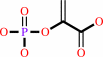 phosphoenolpyruvate
|
+
|
 ADP
ADP
|
+
|
 CO2
CO2
|
|
|
|
|
|
|
|
|
|
|
|
|
Molecule diagrams generated from .mol files obtained from the
KEGG ftp site
|
|
|
|
|
|
|
|
|
|
|
|
|
|
 |
|
|
|
|
|
|
| |
|
|
| |
|
DOI no:
|
J Mol Biol
313:1059-1072
(2001)
|
|
PubMed id:
|
|
|
|
|
|
| |
|
Crystal structure of the dimeric phosphoenolpyruvate carboxykinase (PEPCK) from Trypanosoma cruzi at 2 A resolution.
|
|
S.Trapani,
J.Linss,
S.Goldenberg,
H.Fischer,
A.F.Craievich,
G.Oliva.
|
|
|
|
|
| |
ABSTRACT
|
|
|
|
| |
|
|
ATP-dependent phosphoenolpyruvate carboxykinase (PEPCK) (ATP: oxaloacetate
carboxylyase (transphosphorylating), EC 4.1.1.49) is a key enzyme involved in
the catabolism of glucose and amino acids in the parasite Trypanosoma cruzi, the
causative agent of Chagas' disease. Due to the significant differences in the
amino acid sequence and substrate specificity of the human enzyme (PEPCK
(GTP-dependent), EC 4.1.1.32), the parasite enzyme has been considered a good
target for the development of new anti-chagasic drugs. We have solved the
crystal structure of the recombinant PEPCK of T. cruzi up to 2.0 A resolution,
characterised the dimeric organisation of the enzyme by solution small angle
X-ray scattering (SAXS) and compared the enzyme structure with the known crystal
structure of the monomeric PEPCK from Escherichia coli. The dimeric structure
possesses 2-fold symmetry, with each monomer sharing a high degree of structural
similarity with the monomeric structure of the E. coli PEPCK. Each monomer folds
into two complex mixed alpha/beta domains, with the active site located in a
deep cleft between the domains. The two active sites in the dimer are far apart
from each other, in an arrangement that seems to permit an independent access of
the substrates to the two active sites. All residues of the E. coli PEPCK
structure that had been found to interact with substrates and metal cofactors
have been found conserved and in a substantially equivalent spatial disposition
in the T. cruzi PEPCK structure. No substrate or metal ion was present in the
crystal structure. A sulphate ion from the crystallisation medium has been found
bound to the active site. Solution SAXS data suggest that, in solutions with
lower sulphate concentration than that used for the crystallisation experiments,
the actual enzyme conformation may be slightly different from its conformation
in the crystal structure. This could be due to a conformational transition upon
sulphate binding, similar to the ATP-induced transition observed in the E. coli
PEPCK, or to crystal packing effects. The present structure of the T. cruzi
PEPCK will provide a good basis for the modelling of new anti-chagasic drug
leads.
|
|
|
|
|
|
| |
Selected figure(s)
|
|
|
|
| |
|
|

|
|

|
|
Figure 1.
Figure 1. T. cruzi PEPCK stick model and (3m|F[o]|
-2D|F[c]|) difference Fourier map contoured at 1s level around:
(a) the sulphate ion in the phosphate-binding site; (b) residues
360-379, containing the disordered loop 366-373; the
(incomplete) C^a trace of the corresponding region of the E.
coli PEPCK is shown in green; a discontinuity in the drawing is
due to missing residues in the deposited atomic coordinates of
the E. coli PEPCK. The Figure was drawn using O.[65]
|
|
Figure 7.
Figure 7. The dimeric arrangement of the T. cruzi PEPCK.
N-terminal domains are shown in blue, C-terminal domains in
magenta. The monomer-monomer interface residues are highlighted.
|
|
|
|
|
|
| |
The above figures are
reprinted
by permission from Elsevier:
J Mol Biol
(2001,
313,
1059-1072)
copyright 2001.
|
|
| |
Figures were
selected
by an automated process.
|
|
|
|
|
|
|
|
|
|
|
|
|
|
|
|
 |
 |
 |
|
Literature references that cite this PDB file's key reference
|
|
|
| |
PubMed id
|
|
Reference
|
|
|
|
|
|
E.Pérez,
and
E.Cardemil
(2010).
Saccharomyces cerevisiae phosphoenolpyruvate carboxykinase: the relevance of Glu299 and Leu460 for nucleotide binding.
|
| |
Protein J,
29,
299-305.
|
|
|
|
|
|
|
C.Meesters,
A.Brack,
N.Hellmann,
and
H.Decker
(2009).
Structural characterization of the alpha-hemolysin monomer from Staphylococcus aureus.
|
| |
Proteins,
75,
118-126.
|
|
|
|
|
|
|
G.M.Carlson,
and
T.Holyoak
(2009).
Structural insights into the mechanism of phosphoenolpyruvate carboxykinase catalysis.
|
| |
J Biol Chem,
284,
27037-27041.
|
|
|
|
|
|
|
N.Asanuma,
K.Yoshizawa,
K.Kanada,
and
T.Hino
(2009).
Molecular and biochemical characterization of phosphoenolpyruvate carboxykinase in the ruminal bacterium Ruminococcus albus.
|
| |
Curr Microbiol,
58,
416-420.
|
|
|
|
|
|
|
I.Tobar,
F.D.González-Nilo,
A.M.Jabalquinto,
and
E.Cardemil
(2008).
Relevance of Arg457 for the nucleotide affinity of Saccharomyces cerevisiae phosphoenolpyruvate carboxykinase.
|
| |
Int J Biochem Cell Biol,
40,
1883-1889.
|
|
|
|
|
|
|
A.Yévenes,
F.D.González-Nilo,
and
E.Cardemil
(2007).
Relevance of phenylalanine 216 in the affinity of Saccharomyces cerevisiae phosphoenolpyruvate carboxykinase for Mn(II).
|
| |
Protein J,
26,
135-141.
|
|
|
|
|
|
|
S.Aich,
and
L.T.Delbaere
(2007).
Phylogenetic Study of the Evolution of PEP-Carboxykinase.
|
| |
Evol Bioinform Online,
3,
333-340.
|
|
|
|
|
|
|
A.Takahashi-Terada,
M.Kotera,
K.Ohshima,
T.Furumoto,
H.Matsumura,
Y.Kai,
and
K.Izui
(2005).
Maize phosphoenolpyruvate carboxylase. Mutations at the putative binding site for glucose 6-phosphate caused desensitization and abolished responsiveness to regulatory phosphorylation.
|
| |
J Biol Chem,
280,
11798-11806.
|
|
|
|
|
|
|
J.J.Cotelesage,
L.Prasad,
J.G.Zeikus,
M.Laivenieks,
and
L.T.Delbaere
(2005).
Crystal structure of Anaerobiospirillum succiniciproducens PEP carboxykinase reveals an important active site loop.
|
| |
Int J Biochem Cell Biol,
37,
1829-1837.
|
|
|
PDB codes:
|
|
|
|
|
|
|
|
M.Sugahara,
N.Ohshima,
Y.Ukita,
M.Sugahara,
and
N.Kunishima
(2005).
Structure of ATP-dependent phosphoenolpyruvate carboxykinase from Thermus thermophilus HB8 showing the structural basis of induced fit and thermostability.
|
| |
Acta Crystallogr D Biol Crystallogr,
61,
1500-1507.
|
|
|
PDB codes:
|
|
|
|
|
|
|
|
Y.A.Leduc,
L.Prasad,
M.Laivenieks,
J.G.Zeikus,
and
L.T.Delbaere
(2005).
Structure of PEP carboxykinase from the succinate-producing Actinobacillus succinogenes: a new conserved active-site motif.
|
| |
Acta Crystallogr D Biol Crystallogr,
61,
903-912.
|
|
|
PDB codes:
|
|
|
|
|
|
|
|
C.Bueno,
F.D.González-Nilo,
M.Victoria Encinas,
and
E.Cardemil
(2004).
Substrate binding to fluorescent labeled wild type, Lys213Arg, and HIS233Gln Saccharomyces cerevisiae phosphoenolpyruvate carboxykinases.
|
| |
Int J Biochem Cell Biol,
36,
861-869.
|
|
|
|
|
|
|
S.Aich,
F.Imabayashi,
and
L.T.Delbaere
(2003).
Crystallization and preliminary X-ray crystallographic studies of phosphoenolpyruvate carboxykinase from Corynebacterium glutamicum.
|
| |
Acta Crystallogr D Biol Crystallogr,
59,
1640-1641.
|
|
|
|
|
|
|
M.V.Encinas,
F.D.González-Nilo,
H.Goldie,
and
E.Cardemil
(2002).
Ligand interactions and protein conformational changes of phosphopyridoxyl-labeled Escherichia coli phosphoenolpyruvate carboxykinase determined by fluorescence spectroscopy.
|
| |
Eur J Biochem,
269,
4960-4968.
|
|
|
|
|
|
The most recent references are shown first.
Citation data come partly from CiteXplore and partly
from an automated harvesting procedure. Note that this is likely to be
only a partial list as not all journals are covered by
either method. However, we are continually building up the citation data
so more and more references will be included with time.
Where a reference describes a PDB structure, the PDB
codes are
shown on the right.
|
|


 Links
Links


