Function and Biology

Crystal Structure of ETD97N-EHred complex

Source organism: Escherichia coli K-12
Biochemical function: transferase activity
Biological process: glycine decarboxylation via glycine cleavage system
Cellular component: glycine cleavage complex

EC 2.1.2.10: Aminomethyltransferase

Reaction catalysed:
[Protein]-S(8)-aminomethyldihydrolipoyllysine + tetrahydrofolate = [protein]-dihydrolipoyllysine + 5,10-methylenetetrahydrofolate + NH(3)
Systematic name:
[Protein]-S(8)-aminomethyldihydrolipoyllysine:tetrahydrofolate aminomethyltransferase (ammonia-forming)
Alternative Name(s):
  • Glycine synthase
  • Glycine-cleavage system T-protein
  • S-aminomethyldihydrolipoylprotein:(6S)-tetrahydrofolate aminomethyltransferase (ammonia-forming)
  • T-protein
  • Tetrahydrofolate aminomethyltransferase
  • [Protein]-8-S-aminomethyldihydrolipoyllysine:tetrahydrofolate aminomethyltransferase (ammonia-forming)

Sequence families

Pfam Protein families (Pfam)
PF08669
Domain description: Glycine cleavage T-protein C-terminal barrel domain
Occurring in:
  1. Aminomethyltransferase
The deposited structure of PDB entry 3a8k contains 4 copies of Pfam domain PF08669 (Glycine cleavage T-protein C-terminal barrel domain) in Aminomethyltransferase. Showing 1 copy in chain A.

PF01571
Domain description: Aminomethyltransferase folate-binding domain
Occurring in:
  1. Aminomethyltransferase
The deposited structure of PDB entry 3a8k contains 4 copies of Pfam domain PF01571 (Aminomethyltransferase folate-binding domain) in Aminomethyltransferase. Showing 1 copy in chain A.

PF01597
Domain description: Glycine cleavage H-protein
Occurring in:
  1. Glycine cleavage system H protein
The deposited structure of PDB entry 3a8k contains 2 copies of Pfam domain PF01597 (Glycine cleavage H-protein) in Glycine cleavage system H protein. Showing 1 copy in chain E.

InterPro InterPro annotations
IPR033753
Domain description: Glycine cleavage system H-protein/Simiate
Occurring in:
  1. Glycine cleavage system H protein
IPR002930
Domain description: Glycine cleavage system H-protein
Occurring in:
  1. Glycine cleavage system H protein
IPR013977
Domain description: Glycine cleavage T-protein, C-terminal barrel domain
Occurring in:
  1. Aminomethyltransferase
IPR029043
Domain description: Glycine cleavage T-protein/YgfZ, C-terminal
Occurring in:
  1. Aminomethyltransferase
IPR006222
Domain description: Aminomethyltransferase, folate-binding domain
Occurring in:
  1. Aminomethyltransferase
IPR027266
Domain description: GTP-binding protein TrmE/Aminomethyltransferase GcvT, domain 1
Occurring in:
  1. Aminomethyltransferase
IPR028896
Domain description: Aminomethyltransferase-like
Occurring in:
  1. Aminomethyltransferase
IPR006223
Domain description: Glycine cleavage system T protein
Occurring in:
  1. Aminomethyltransferase
IPR022903
Domain description: Glycine cleavage system T protein, bacteria
Occurring in:
  1. Aminomethyltransferase
IPR003016
Domain description: 2-oxo acid dehydrogenase, lipoyl-binding site
Occurring in:
  1. Glycine cleavage system H protein
IPR000089
Domain description: Biotin/lipoyl attachment
Occurring in:
  1. Glycine cleavage system H protein
IPR011053
Domain description: Single hybrid motif
Occurring in:
  1. Glycine cleavage system H protein
IPR017453
Domain description: Glycine cleavage system H-protein, subgroup
Occurring in:
  1. Glycine cleavage system H protein

Structure domains

CATH CATH domains
3.30.1360.120
Class: Alpha Beta
Architecture: 2-Layer Sandwich
Topology: Gyrase A; domain 2
Homology: Probable tRNA modification gtpase trme; domain 1
Occurring in:
  1. Aminomethyltransferase
The deposited structure of PDB entry 3a8k contains 4 copies of CATH domain 3.30.1360.120 (Gyrase A; domain 2) in Aminomethyltransferase. Showing 1 copy in chain A.
2.40.30.110
Class: Mainly Beta
Architecture: Beta Barrel
Topology: Elongation Factor Tu (Ef-tu); domain 3
Homology: Aminomethyltransferase beta-barrel domains
Occurring in:
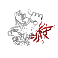
  1. Aminomethyltransferase
The deposited structure of PDB entry 3a8k contains 4 copies of CATH domain 2.40.30.110 (Elongation Factor Tu (Ef-tu); domain 3) in Aminomethyltransferase. Showing 1 copy in chain A.
4.10.1250.10
Class: Few Secondary Structures
Architecture: Irregular
Topology: Aminomethyltransferase fragment
Homology: Aminomethyltransferase fragment
Occurring in:
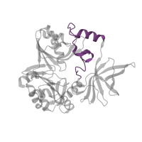
  1. Aminomethyltransferase
The deposited structure of PDB entry 3a8k contains 4 copies of CATH domain 4.10.1250.10 (Aminomethyltransferase fragment) in Aminomethyltransferase. Showing 1 copy in chain A.
3.30.70.1400
Class: Alpha Beta
Architecture: 2-Layer Sandwich
Topology: Alpha-Beta Plaits
Homology: Aminomethyltransferase beta-barrel domains
Occurring in:
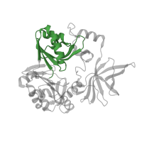
  1. Aminomethyltransferase
The deposited structure of PDB entry 3a8k contains 4 copies of CATH domain 3.30.70.1400 (Alpha-Beta Plaits) in Aminomethyltransferase. Showing 1 copy in chain A.
2.40.50.100
Class: Mainly Beta
Architecture: Beta Barrel
Topology: OB fold (Dihydrolipoamide Acetyltransferase, E2P)
Homology: OB fold (Dihydrolipoamide Acetyltransferase, E2P)
Occurring in:
  1. Glycine cleavage system H protein
The deposited structure of PDB entry 3a8k contains 2 copies of CATH domain 2.40.50.100 (OB fold (Dihydrolipoamide Acetyltransferase, E2P)) in Glycine cleavage system H protein. Showing 1 copy in chain E.