EMD-9876
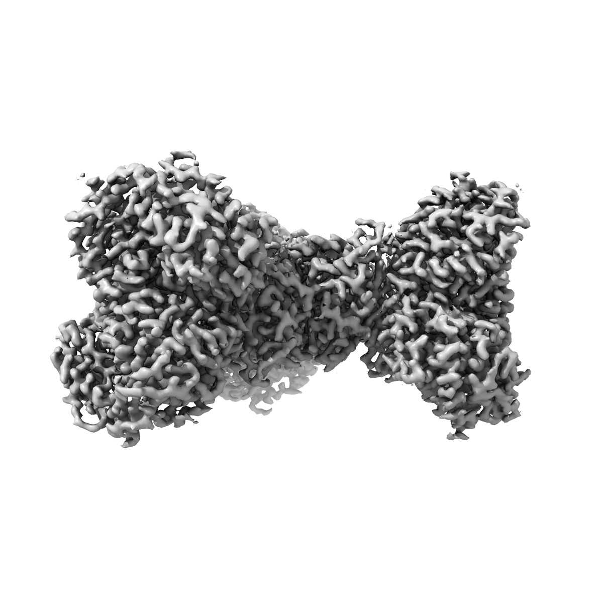
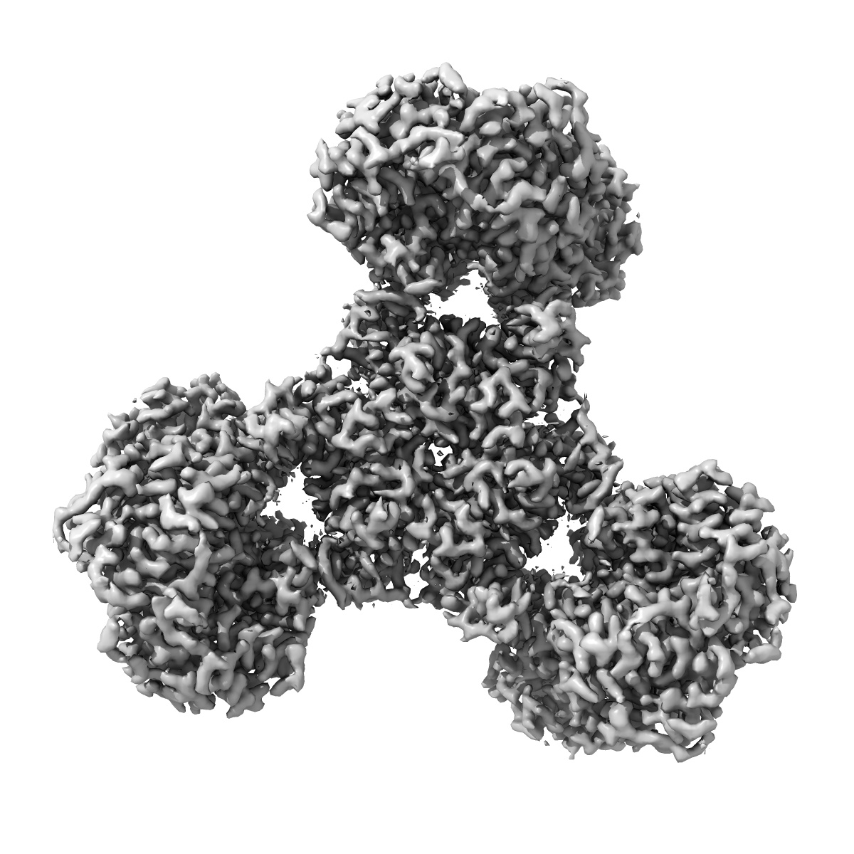
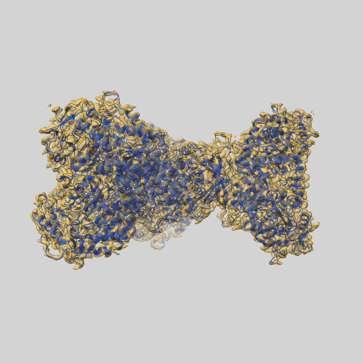
Structure of PaaZ, a bifunctional enzyme in complex with NADP+ and CCoA
EMD-9876
Single-particle3.1 Å
 Deposition: 31/03/2019
Map released: 11/09/2019
Last modified: 27/03/2024
Concentration: 0.015
mg/mL
Details: The peak fraction from gel filtration was used for grid preparation. NADP+ and CCoA were added 10 fold excess and incubated for 15 minutes before applying to the grid.
Details: The peak fraction from gel filtration was used for grid preparation. NADP+ and CCoA were added 10 fold excess and incubated for 15 minutes before applying to the grid.
Buffer
Grid
Mesh: 300
Model: Quantifoil, UltrAuFoil, R1.2/1.3
Material: GOLD
Details: Grids were glow discharged for 5 minutes and graphene oxide was applied. This was followed by 3 times wash with water and dried.
Model: Quantifoil, UltrAuFoil, R1.2/1.3
Material: GOLD
Details: Grids were glow discharged for 5 minutes and graphene oxide was applied. This was followed by 3 times wash with water and dried.
Pretreatment
Support Film [1]
| Material | Topology | Thickness |
|---|---|---|
| GRAPHENE OXIDE | CONTINUOUS | - |
Vitrification
Cryogen name: ETHANE
Chamber humidity: 100%
Chamber temperature: 283.15 K
Instrument: FEI VITROBOT MARK IV
Details: Blotting force 10, blotting time 4 sec, waiting time 15 sec, drying time 0, blotting time 1..
Chamber humidity: 100%
Chamber temperature: 283.15 K
Instrument: FEI VITROBOT MARK IV
Details: Blotting force 10, blotting time 4 sec, waiting time 15 sec, drying time 0, blotting time 1..
Microscope: FEI TITAN KRIOS
Illumination mode: FLOOD BEAM
Imaging mode: BRIGHT FIELD
Electron source: FIELD EMISSION GUN
Acceleration voltage: 300 kV
C2 aperture diameter: 50.0 µm
Nominal CS: 2.7 mm
Nominal defocus: 2.2 µm - 3.2 µm
Nominal magnification: 75000.0
Calibrated magnification: 134615.0
Specimen holder model: FEI TITAN KRIOS AUTOGRID HOLDER
Cooling holder cryogen: NITROGEN
Alignment procedure: COMA FREE
Details: Data was collected with EPU software
Temperature
Minimum: 80.0
K
Maximum: 80.0 K
Maximum: 80.0 K
Image Recording
[1]
Detector model:
FEI FALCON III (4k x 4k)
Detector mode: COUNTING
Dimensions: 4096 pixel x 4096 pixel
Number of grids: 1
Number of real images: 563
Average exposure time: 60.0 s
Average electron dose per image: 27.0 e/Å2
Details: The 60 second exposure was saved into 75 frames with each frame ~0.36 e-. The frames were then grouped into 3 for alignment and summed images were used for data processing
Details: Counting mode
Final
reconstruction
Resolution: 3.1
Å
(
BY AUTHOR)
Resolution method: FSC 0.143 CUT-OFF
Number of classed used: 1
Number of images used: 120968
Algorithm: BACK PROJECTION
Details:
Resolution method: FSC 0.143 CUT-OFF
Number of classed used: 1
Number of images used: 120968
Algorithm: BACK PROJECTION
Details:
⌯ Applied Symmetry
Point group:
C3
Software
[1]
| Name | Version | Details |
|---|---|---|
| RELION | 2.0 | - |
Startup model
[1]
⦨ Initial angle
assignment
⦩ Final angle assignment
Particle selection
[1]
| Selected | Ref. model | Method | Software | Details |
|---|---|---|---|---|
| 122910 | - | - | - | - |
Final 3D classification
Software
[1]
| Name | Version | Details |
|---|---|---|
| RELION | 2.0 | - |
Format: CCP4
Data type: IMAGE STORED AS FLOATING POINT NUMBER (4 BYTES)
Annotation details: The map of PaaZ is in complex with NADP and Crotonyl CoA
Data type: IMAGE STORED AS FLOATING POINT NUMBER (4 BYTES)
Annotation details: The map of PaaZ is in complex with NADP and Crotonyl CoA
⬡ Geometry
| X | Y | Z | |
|---|---|---|---|
| Dimensions | 412 | 412 | 412 |
| Origin | 0 | 0 | 0 |
| Spacing | 412 | 412 | 412 |
| Voxel size | 1.04 Å | 1.04 Å | 1.04 Å |
Contour list
| Primary | Level | Source |
|---|---|---|
| True | 0.085 | AUTHOR |