EMD-9843
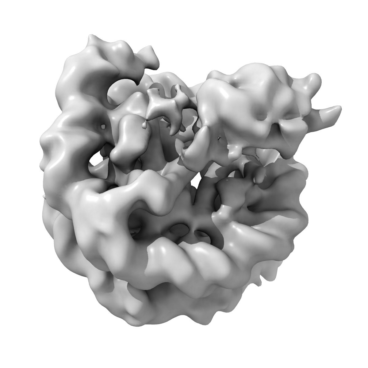
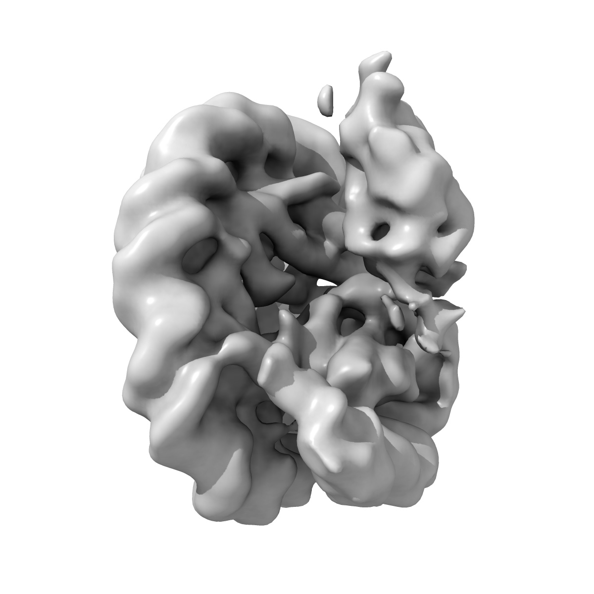
cryo-EM structure of DOT1L bound to unmodified nucleosome
EMD-9843
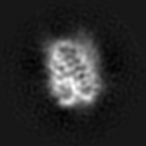
Single-particle7.3 Å
 Deposition: 07/03/2019
Map released: 15/05/2019
Last modified: 27/03/2024
Buffer
pH: 7.5
Vitrification
Cryogen name: ETHANE
Microscope: FEI TITAN
Illumination mode: OTHER
Imaging mode: BRIGHT FIELD
Electron source: FIELD EMISSION GUN
Acceleration voltage: 300 kV
Illumination mode: OTHER
Imaging mode: BRIGHT FIELD
Electron source: FIELD EMISSION GUN
Acceleration voltage: 300 kV
Image Recording
[1]
Final
reconstruction
Startup model
[1]
Type:
OTHER
⦨ Initial angle
assignment
Type:
MAXIMUM LIKELIHOOD
⦩ Final angle assignment
Type:
MAXIMUM LIKELIHOOD
Format: CCP4
Data type: IMAGE STORED AS FLOATING POINT NUMBER (4 BYTES)
Annotation details: Structure of catalytic domain of DOT1L bound to core nucleosome
Data type: IMAGE STORED AS FLOATING POINT NUMBER (4 BYTES)
Annotation details: Structure of catalytic domain of DOT1L bound to core nucleosome
⬡ Geometry
| X | Y | Z | |
|---|---|---|---|
| Dimensions | 200 | 200 | 200 |
| Origin | 0 | 0 | 0 |
| Spacing | 200 | 200 | 200 |
| Voxel size | 1.06 Å | 1.06 Å | 1.06 Å |
Contour list
| Primary | Level | Source |
|---|---|---|
| True | 0.021 | AUTHOR |