EMD-9232
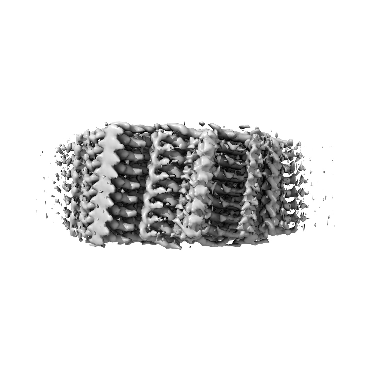
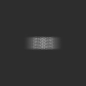
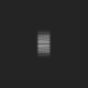
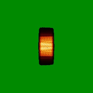
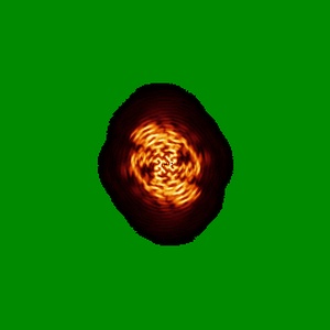
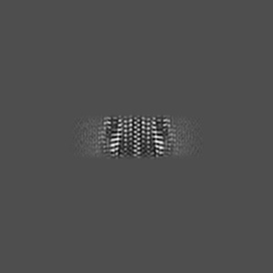
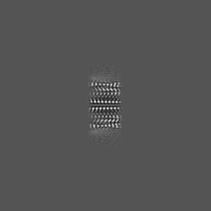
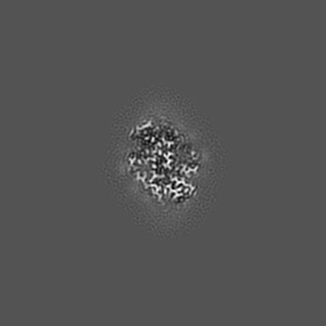
Cryo-EM structure of human AA amyloid fibril
EMD-9232
Helical reconstruction2.7 Å
 Deposition: 18/10/2018
Map released: 13/03/2019
Last modified: 13/03/2024
Concentration: 0.2
mg/mL
Buffer
pH: 7.0
Buffer components [1]:
Buffer components [1]:
| Name | Formula | Concentration | ChEBI |
|---|---|---|---|
| - | ddH2O | - |
Grid
Vitrification
Cryogen name: ETHANE
Chamber humidity: 95%
Chamber temperature: 293 K
Instrument: FEI VITROBOT MARK III
Chamber humidity: 95%
Chamber temperature: 293 K
Instrument: FEI VITROBOT MARK III
Microscope: FEI TITAN KRIOS
Illumination mode: FLOOD BEAM
Imaging mode: BRIGHT FIELD
Electron source: FIELD EMISSION GUN
Acceleration voltage: 300 kV
C2 aperture diameter: 50.0 µm
Nominal CS: 2.7 mm
Nominal defocus: -0.5 µm - -2.5 µm
Nominal magnification: 130000.0
Cooling holder cryogen: NITROGEN
Illumination mode: FLOOD BEAM
Imaging mode: BRIGHT FIELD
Electron source: FIELD EMISSION GUN
Acceleration voltage: 300 kV
C2 aperture diameter: 50.0 µm
Nominal CS: 2.7 mm
Nominal defocus: -0.5 µm - -2.5 µm
Nominal magnification: 130000.0
Cooling holder cryogen: NITROGEN
Image Recording
[1]
Detector model:
GATAN K2 SUMMIT (4k x 4k)
Detector mode: COUNTING
Average exposure time: 12.0 s
Average electron dose per image: 40.0 e/Å2
Detector mode: COUNTING
Average exposure time: 12.0 s
Average electron dose per image: 40.0 e/Å2
Final
reconstruction
Resolution: 2.7
Å
(
BY AUTHOR)
Resolution method: FSC 0.143 CUT-OFF
Number of images used: 91872
Resolution method: FSC 0.143 CUT-OFF
Number of images used: 91872
⌯ Applied Symmetry
Software
[1]
| Name | Version | Details |
|---|---|---|
| RELION | 2.1 | - |
Startup model
[1]
Type:
NONE
⦩ Final angle assignment
Segment selection
[1]
| Number selected | Segment length | Segment overlap | Total filament length | Details |
|---|---|---|---|---|
| 93025 | - | - | - | - |
Format: CCP4
Data type: IMAGE STORED AS FLOATING POINT NUMBER (4 BYTES)
Annotation details: Cryo-EM reconstruction of human Serum Amyloid A fibrils, extracted from diseased human kidney. The fibril shows a helical rise of 2.40 A, a helical twist of 180.79 degree and a resolution of 2.7 A.
Data type: IMAGE STORED AS FLOATING POINT NUMBER (4 BYTES)
Annotation details: Cryo-EM reconstruction of human Serum Amyloid A fibrils, extracted from diseased human kidney. The fibril shows a helical rise of 2.40 A, a helical twist of 180.79 degree and a resolution of 2.7 A.
⬡ Geometry
| X | Y | Z | |
|---|---|---|---|
| Dimensions | 270 | 270 | 270 |
| Origin | 0 | 0 | 0 |
| Spacing | 270 | 270 | 270 |
| Voxel size | 1.04 Å | 1.04 Å | 1.04 Å |
Contour list
| Primary | Level | Source |
|---|---|---|
| True | 0.05 | AUTHOR |