EMD-8215
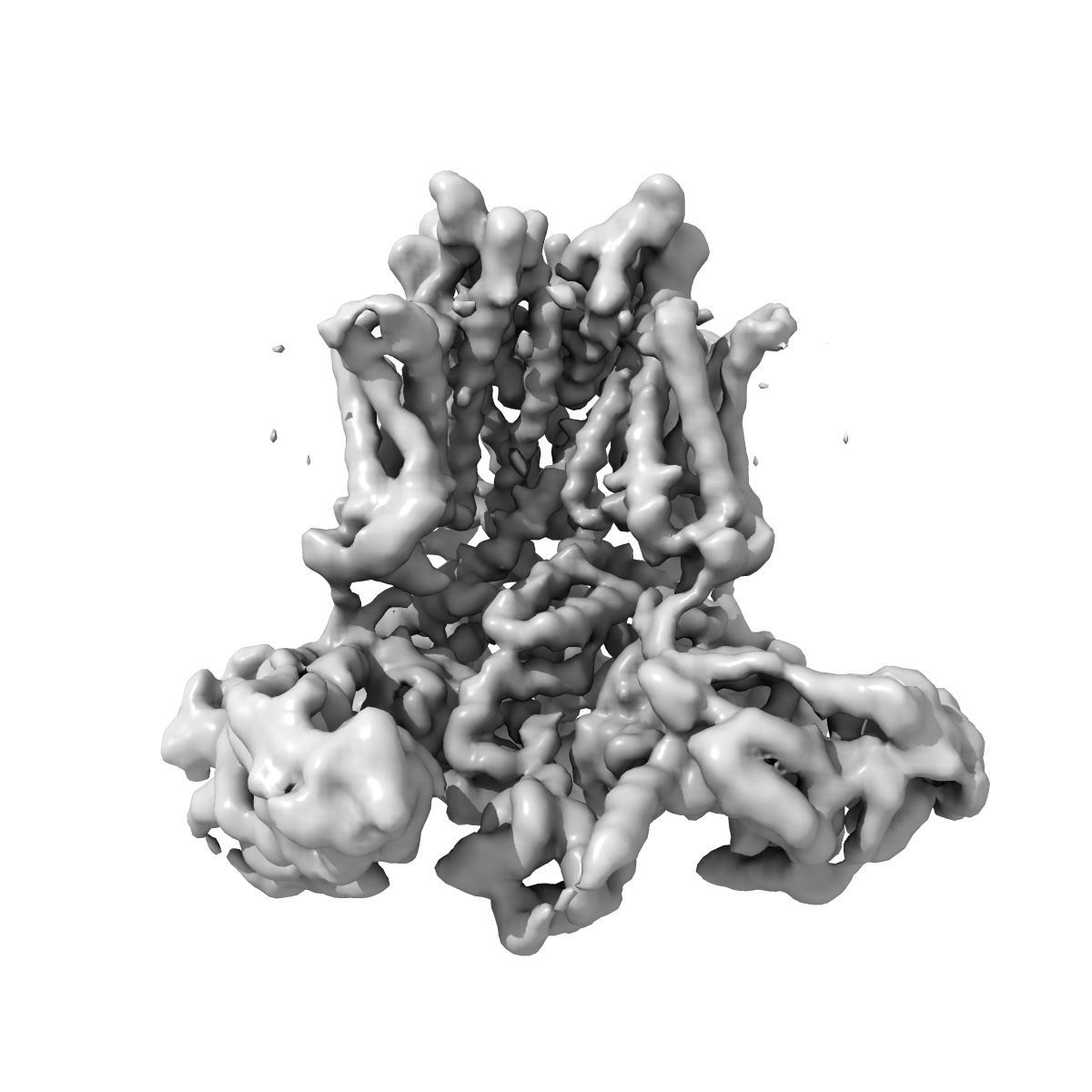
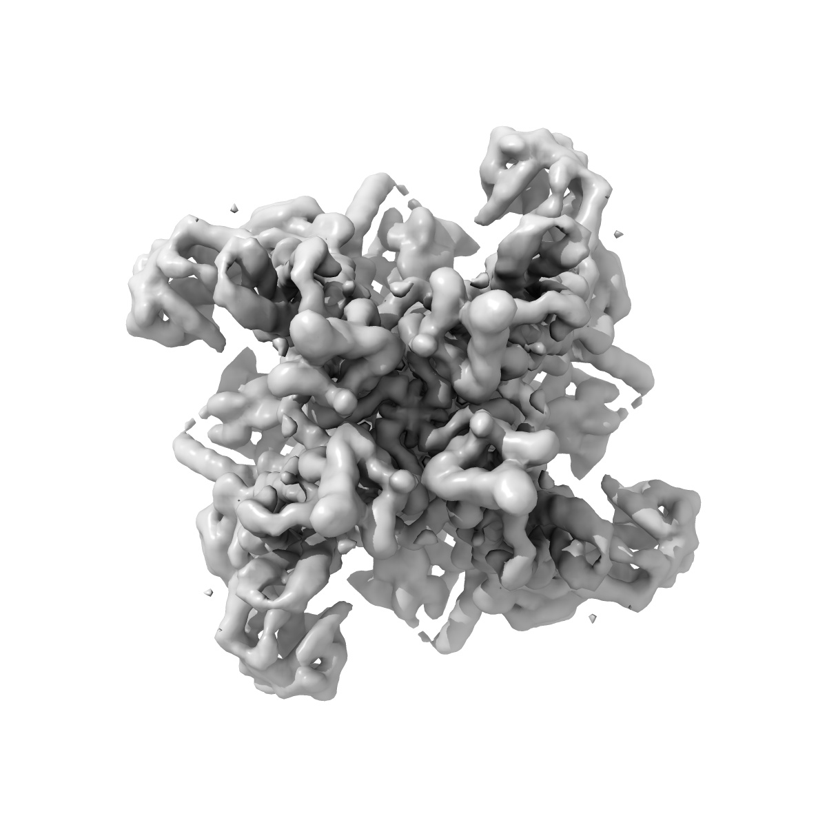
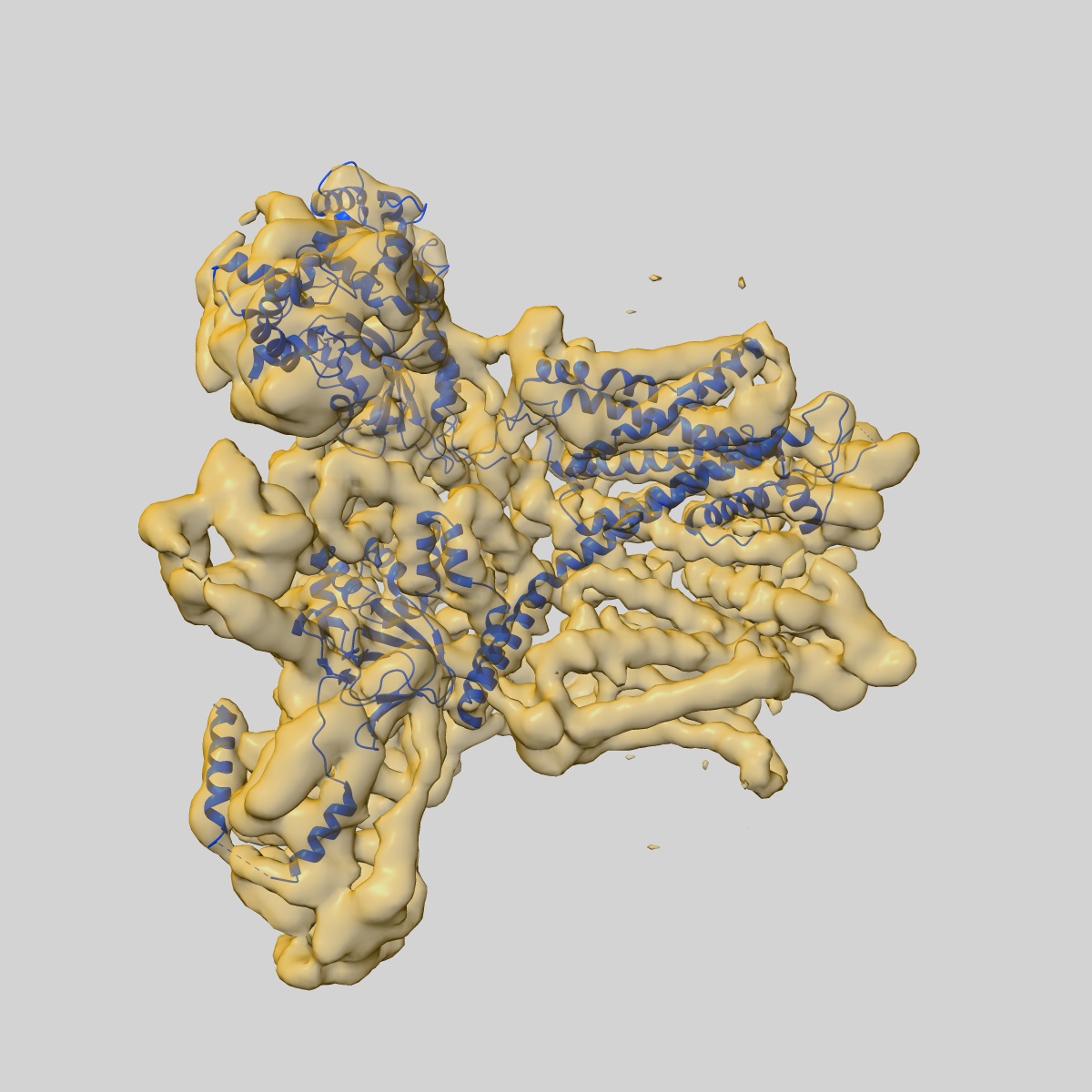
Single particle cryo-EM structure of the voltage-gated K+ channel Eag1 bound to the channel inhibitor calmodulin
EMD-8215
Single-particle3.78 Å
 Deposition: 16/06/2016
Map released: 17/08/2016
Last modified: 29/07/2020
Name: Voltage-gated K+ channel Eag1 bound to the channel inhibitor calmodulin
Summary:
Name: Voltage-gated K+ channel Eag1 bound to the channel inhibitor calmodulin
Molecular weight
| Experimental | Theoretical | Method |
|---|---|---|
| - | 450 kDa | - |
Name: Voltage-gated potassium channel Eag1
Natural source [1]
Recombinant Expression [4]
| Organism | Strain | Cell | Plasmid |
|---|---|---|---|
| - | - | - | - |
| - | - | - | - |
| - | - | - | - |
| - | - | - | - |
Molecular weight
| Experimental | Theoretical | Method |
|---|---|---|
| - | 380 kDa | - |
Name: Calmodulin
Natural source [1]
Organism: Homo sapiens
Recombinant Expression [4]
| Organism | Strain | Cell | Plasmid |
|---|---|---|---|
| - | - | - | - |
| - | - | - | - |
| - | - | - | - |
| - | - | - | - |
Molecular weight
| Experimental | Theoretical | Method |
|---|---|---|
| - | 17 kDa | - |
Name: Potassium voltage-gated channel subfamily H member 1
Number of copies: 1
UniProtKB PDBe-KB AlphaFold DB
Number of copies: 1
UniProtKB PDBe-KB AlphaFold DB
Domains
Gene Ontology [3]
Biological process:
potassium ion transmembrane transport
Molecular function:
voltage-gated potassium channel activity
Cellular location:
integral component of plasma membrane
potassium ion transmembrane transport
Molecular function:
voltage-gated potassium channel activity
Cellular location:
integral component of plasma membrane
Natural source
Organism: Rattus norvegicus
Molecular weight
| Experimental | Theoretical | Method |
|---|---|---|
| - | 97 kDa | - |
Recombinant Expression
| Organism | Strain | Cell | Plasmid |
|---|---|---|---|
| Homo sapiens | - | - | - |
Name: Calmodulin
Number of copies: 1
UniProtKB PDBe-KB AlphaFold DB
Number of copies: 1
UniProtKB PDBe-KB AlphaFold DB
Domains
Gene Ontology [63]
Biological process:
response to calcium ion cellular response to interferon-beta positive regulation of receptor signaling pathway via JAK-STAT cellular response to type II interferon regulation of calcium-mediated signaling autophagosome membrane docking positive regulation of protein autophosphorylation positive regulation of peptidyl-threonine phosphorylation negative regulation of calcium ion export across plasma membrane substantia nigra development positive regulation of DNA binding regulation of heart rate positive regulation of protein dephosphorylation regulation of cytokinesis regulation of ryanodine-sensitive calcium-release channel activity positive regulation of cyclic-nucleotide phosphodiesterase activity positive regulation of ryanodine-sensitive calcium-release channel activity positive regulation of protein serine/threonine kinase activity regulation of cell communication by electrical coupling involved in cardiac conduction G2/M transition of mitotic cell cycle negative regulation of ryanodine-sensitive calcium-release channel activity regulation of cardiac muscle contraction by regulation of the release of sequestered calcium ion G protein-coupled receptor signaling pathway regulation of cardiac muscle cell action potential negative regulation of peptidyl-threonine phosphorylation regulation of release of sequestered calcium ion into cytosol by sarcoplasmic reticulum mitochondrion-endoplasmic reticulum membrane tethering positive regulation of phosphoprotein phosphatase activity organelle localization by membrane tethering negative regulation of high voltage-gated calcium channel activity detection of calcium ion regulation of cardiac muscle contraction
Molecular function:
enzyme regulator activity calcium ion binding adenylate cyclase activator activity N-terminal myristoylation domain binding calcium-dependent protein binding adenylate cyclase binding protein phosphatase activator activity protein domain specific binding protein serine/threonine kinase activator activity calcium channel inhibitor activity transmembrane transporter binding disordered domain specific binding titin binding protein kinase binding
Cellular location:
sperm midpiece extracellular region cytosol centrosome nucleus myelin sheath plasma membrane spindle pole sarcomere vesicle spindle microtubule calcium channel complex catalytic complex voltage-gated potassium channel complex protein-containing complex nucleoplasm cytoplasm
Natural source
Organism: Homo sapiens
Molecular weight
| Experimental | Theoretical | Method |
|---|---|---|
| - | 16 kDa | - |
Recombinant Expression
| Organism | Strain | Cell | Plasmid |
|---|---|---|---|
| Homo sapiens | - | - | - |
Name: 2-acetamido-2-deoxy-beta-D-glucopyranose
HET code: NAG
Number of copies: 1
ChEMBL ChEBI
HET code: NAG
Number of copies: 1
ChEMBL ChEBI
Molecular weight
| Experimental | Theoretical | Method |
|---|---|---|
| - | 221 Da | - |
Name: CHOLESTEROL HEMISUCCINATE
HET code: Y01
Number of copies: 1
ChEBI
HET code: Y01
Number of copies: 1
ChEBI
Molecular weight
| Experimental | Theoretical | Method |
|---|---|---|
| - | 486 Da | - |