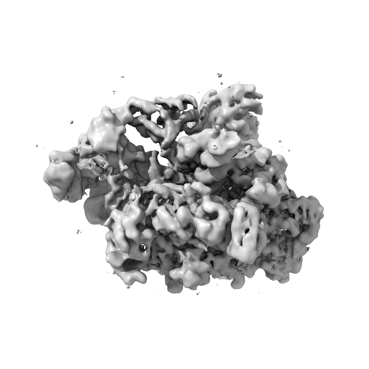
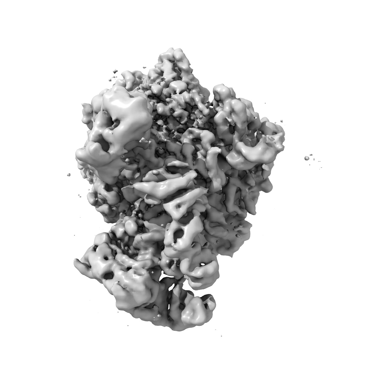
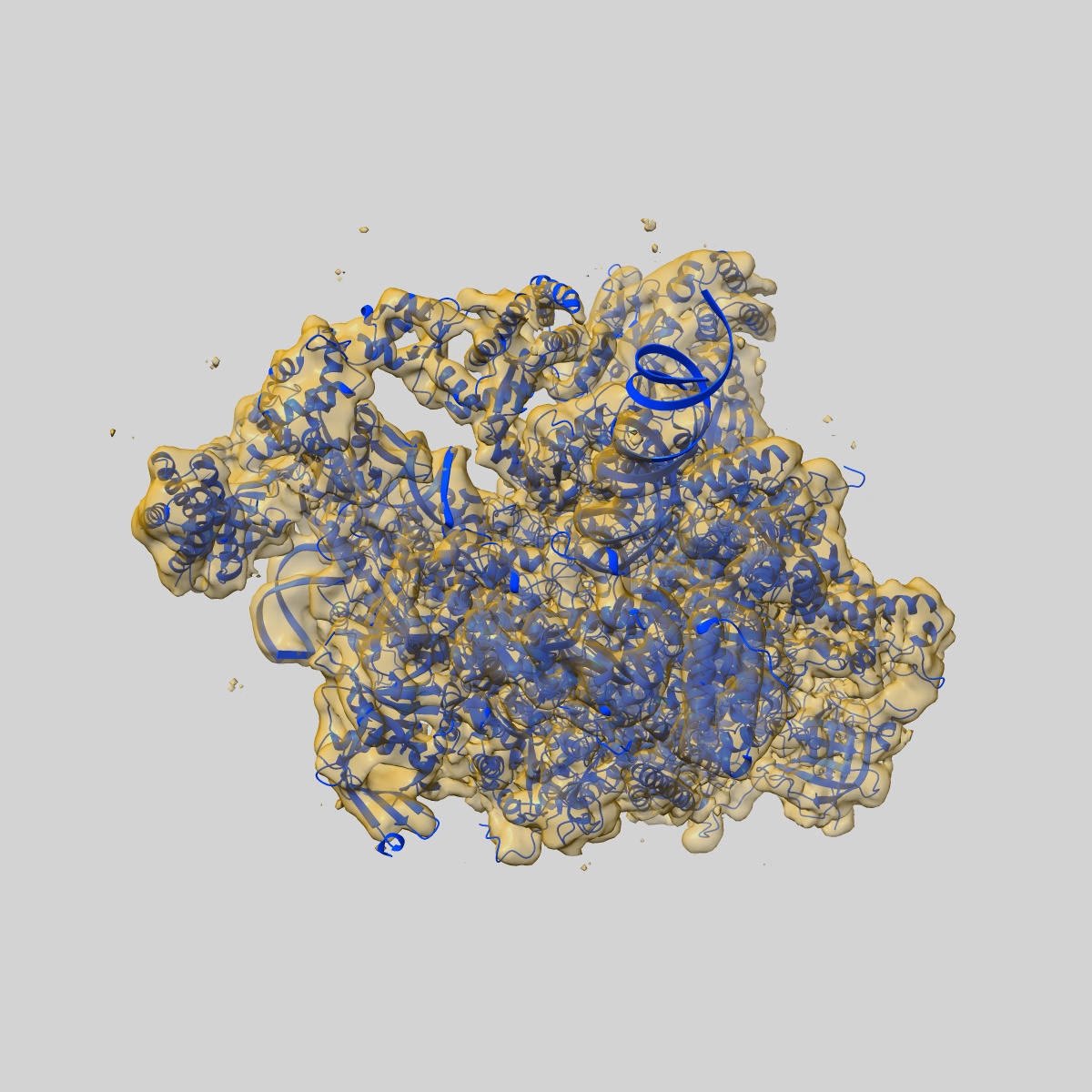
EMD-8136
Human core-PIC in the open state
EMD-8136
Single-particle3.9 Å
 Deposition: 02/05/2016
Map released: 18/05/2016
Last modified: 06/03/2024
Concentration: 0.3
mg/mL
Buffer
pH: 7.9
Vitrification
Cryogen name: ETHANE
Chamber humidity: 100%
Instrument: FEI VITROBOT MARK IV
Details: Blot for 4 seconds before plunging into liquid ethane (FEI VITROBOT MARK IV)..
Chamber humidity: 100%
Instrument: FEI VITROBOT MARK IV
Details: Blot for 4 seconds before plunging into liquid ethane (FEI VITROBOT MARK IV)..
Microscope: FEI TITAN KRIOS
Illumination mode: FLOOD BEAM
Imaging mode: BRIGHT FIELD
Electron source: FIELD EMISSION GUN
Acceleration voltage: 300 kV
Nominal CS: 2.7 mm
Nominal defocus: 2.0 µm - 4.0 µm
Nominal magnification: 27500.0
Specimen holder model: GATAN 626 SINGLE TILT LIQUID NITROGEN CRYO TRANSFER HOLDER
Illumination mode: FLOOD BEAM
Imaging mode: BRIGHT FIELD
Electron source: FIELD EMISSION GUN
Acceleration voltage: 300 kV
Nominal CS: 2.7 mm
Nominal defocus: 2.0 µm - 4.0 µm
Nominal magnification: 27500.0
Specimen holder model: GATAN 626 SINGLE TILT LIQUID NITROGEN CRYO TRANSFER HOLDER
Image Recording
[1]
Detector model:
GATAN K2 SUMMIT (4k x 4k)
Detector mode: COUNTING
Average electron dose per image: 42.0 e/Å2
Detector mode: COUNTING
Average electron dose per image: 42.0 e/Å2
Final
reconstruction
Resolution: 3.9
Å
(
BY AUTHOR)
Resolution method: FSC 0.143 CUT-OFF
Number of images used: 79849
Resolution method: FSC 0.143 CUT-OFF
Number of images used: 79849
Software
[1]
| Name | Version | Details |
|---|---|---|
| RELION | 1.4beta | - |
Startup model
[1]
Type:
OTHER
Details:Negative-stain reconstruction of a similar complex, low-pass filtered to 60 Angstrom resolution
Details:Negative-stain reconstruction of a similar complex, low-pass filtered to 60 Angstrom resolution
⦨ Initial angle
assignment
Type:
PROJECTION MATCHING
⦩ Final angle assignment
Type:
PROJECTION MATCHING
Format: CCP4
Data type: IMAGE STORED AS FLOATING POINT NUMBER (4 BYTES)
Annotation details: None
Data type: IMAGE STORED AS FLOATING POINT NUMBER (4 BYTES)
Annotation details: None
⬡ Geometry
| X | Y | Z | |
|---|---|---|---|
| Dimensions | 384 | 384 | 384 |
| Origin | 0 | 0 | 0 |
| Spacing | 384 | 384 | 384 |
| Voxel size | 1.31 Å | 1.31 Å | 1.31 Å |
Contour list
| Primary | Level | Source |
|---|---|---|
| True | 0.01 | AUTHOR |