EMD-7780
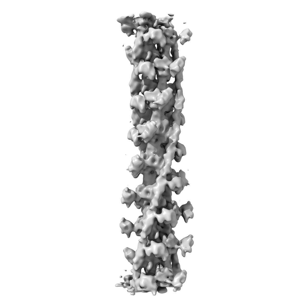
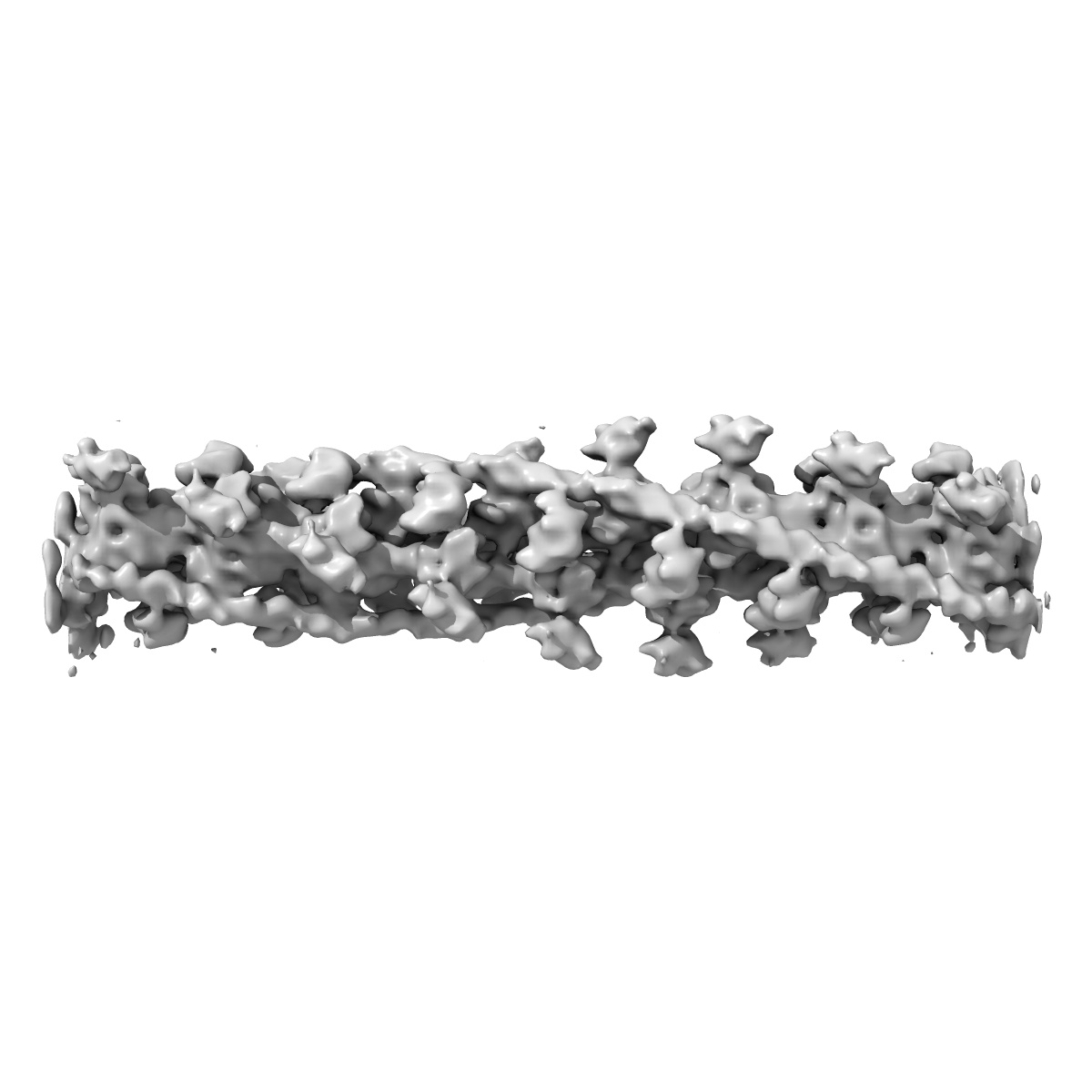
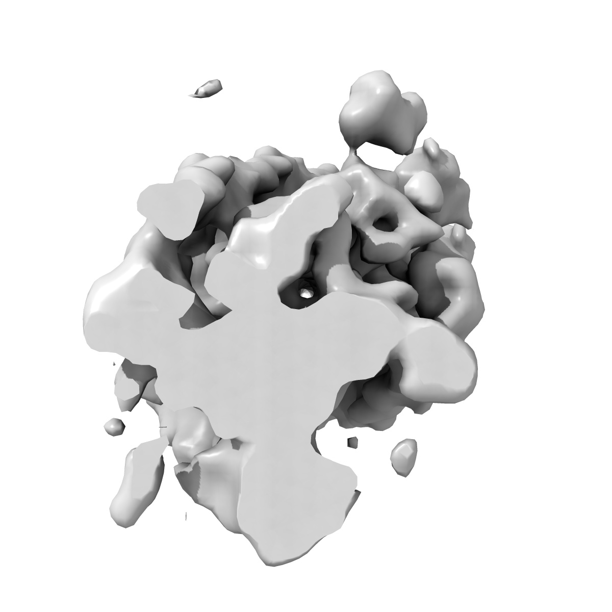
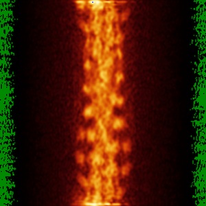
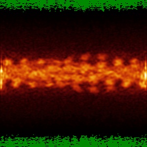
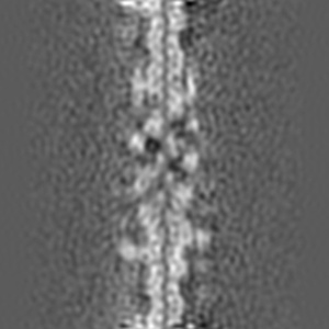
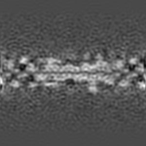
Cardiac thin filament decorated with C0C1 fragment of cardiac myosin binding protein C mode 1
EMD-7780
Helical reconstruction11.0 Å
 Deposition: 03/04/2018
Map released: 10/10/2018
Last modified: 13/03/2024
Buffer
pH: 7.0
Grid
Vitrification
Cryogen name: ETHANE
Chamber humidity: 95%
Chamber temperature: 294 K
Instrument: FEI VITROBOT MARK II
Chamber humidity: 95%
Chamber temperature: 294 K
Instrument: FEI VITROBOT MARK II
Microscope: FEI TITAN KRIOS
Illumination mode: FLOOD BEAM
Imaging mode: BRIGHT FIELD
Electron source: FIELD EMISSION GUN
Acceleration voltage: 300 kV
Illumination mode: FLOOD BEAM
Imaging mode: BRIGHT FIELD
Electron source: FIELD EMISSION GUN
Acceleration voltage: 300 kV
Image Recording
[1]
Detector model:
FEI FALCON II (4k x 4k)
Detector mode: INTEGRATING
Average electron dose per image: 20.0 e/Å2
Detector mode: INTEGRATING
Average electron dose per image: 20.0 e/Å2
Final
reconstruction
Resolution: 11.0
Å
(
BY AUTHOR)
Resolution method: FSC 0.143 CUT-OFF
Number of images used: 6117
Algorithm: BACK PROJECTION
Resolution method: FSC 0.143 CUT-OFF
Number of images used: 6117
Algorithm: BACK PROJECTION
⌯ Applied Symmetry
Software
[1]
| Name | Version | Details |
|---|---|---|
| SPIDER | - | IHRSR |
Startup model
[1]
⦩ Final angle assignment
Format: CCP4
Data type: IMAGE STORED AS FLOATING POINT NUMBER (4 BYTES)
Annotation details: Cardiac thin filament decorated with C0C1 fragment of cardiac myosin binding protein C
Data type: IMAGE STORED AS FLOATING POINT NUMBER (4 BYTES)
Annotation details: Cardiac thin filament decorated with C0C1 fragment of cardiac myosin binding protein C
⬡ Geometry
| X | Y | Z | |
|---|---|---|---|
| Dimensions | 230 | 230 | 230 |
| Origin | -115 | -115 | -114 |
| Spacing | 230 | 230 | 230 |
| Voxel size | 2.1 Å | 2.1 Å | 2.1 Å |
Contour list
| Primary | Level | Source |
|---|---|---|
| True | 5304.0 | AUTHOR |