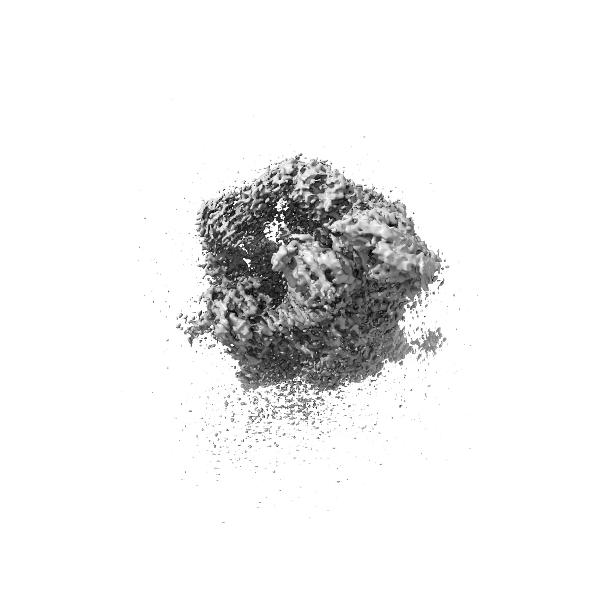
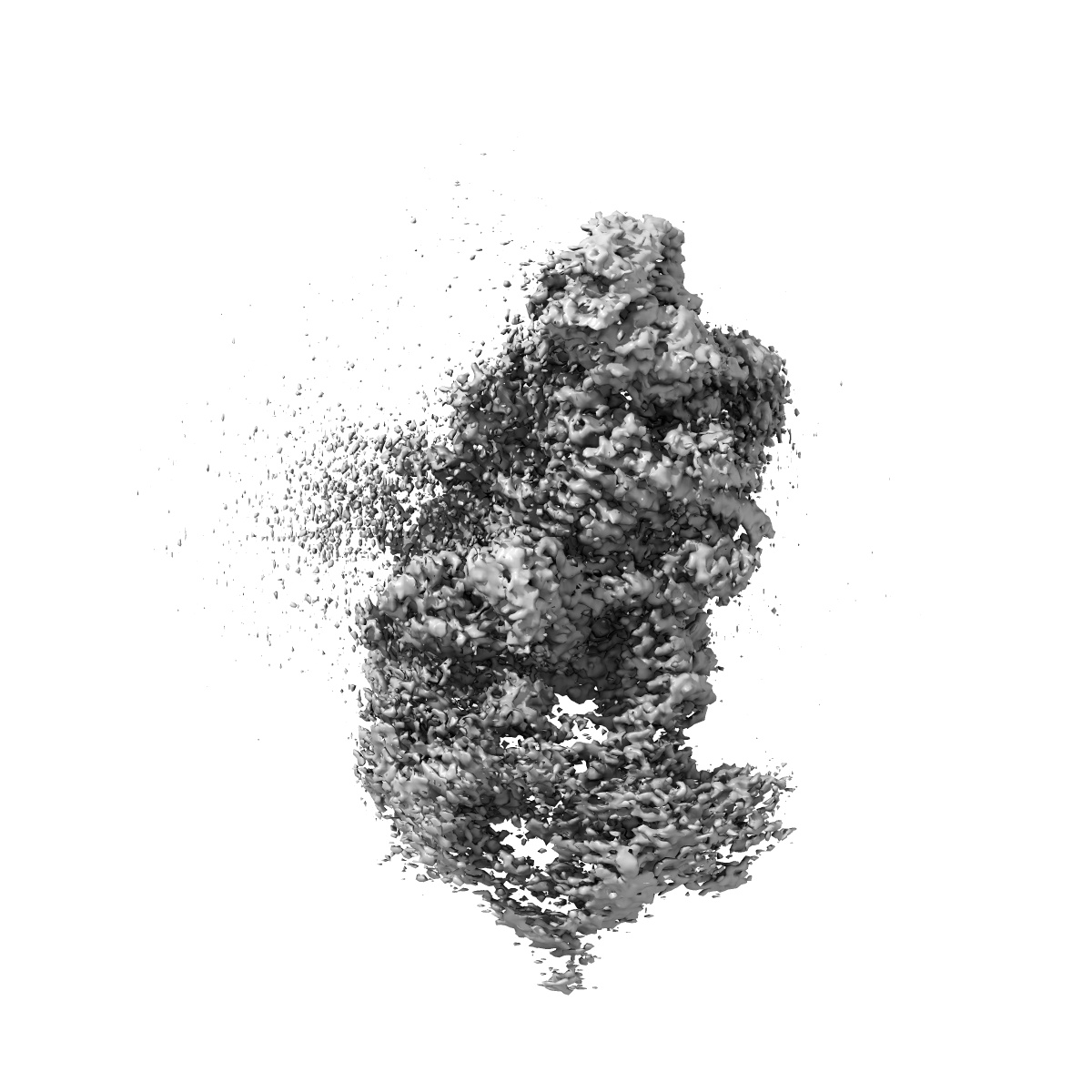
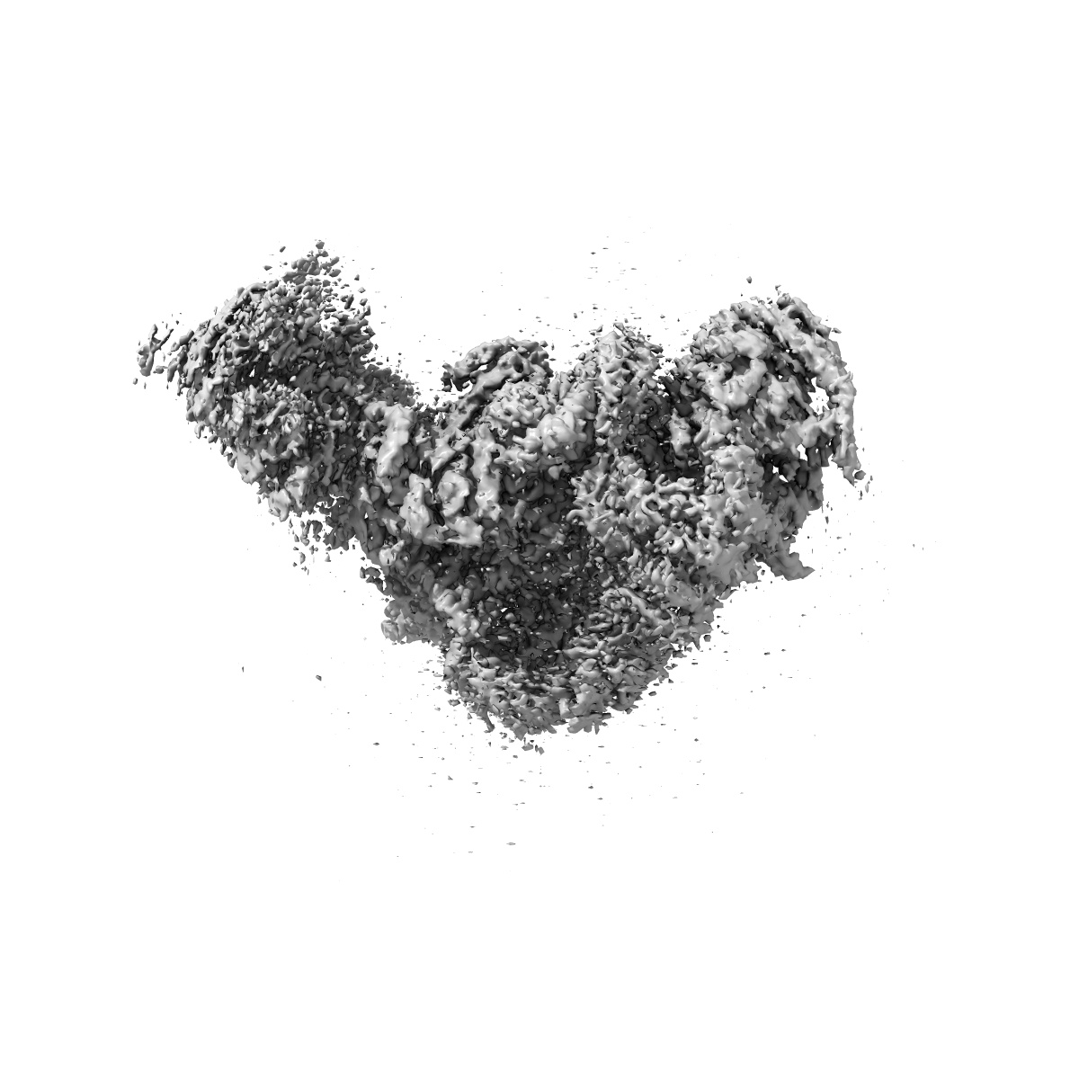
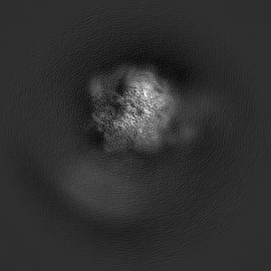
EMD-6566
The cryo-EM structure of yeast spliceosomal U4/U6.U5 tri-snRNP (improved map for Prp3 region at 3.46 A)
EMD-6566
Single-particle3.46 Å
 Deposition: 23/12/2015
Map released: 10/02/2016
Last modified: 10/02/2016
Vitrification
Microscope: FEI TITAN KRIOS
Illumination mode: FLOOD BEAM
Imaging mode: BRIGHT FIELD
Electron source: FIELD EMISSION GUN
Acceleration voltage: 300 kV
Specimen holder model: FEI TITAN KRIOS AUTOGRID HOLDER
Illumination mode: FLOOD BEAM
Imaging mode: BRIGHT FIELD
Electron source: FIELD EMISSION GUN
Acceleration voltage: 300 kV
Specimen holder model: FEI TITAN KRIOS AUTOGRID HOLDER
Image Recording
[1]
Detector category:
FILM
Detector model: GATAN K2 SUMMIT (4k x 4k)
Scanner: TEMSCAN
Number of real images: 3141
Detector model: GATAN K2 SUMMIT (4k x 4k)
Scanner: TEMSCAN
Number of real images: 3141
Format: CCP4
Data type: IMAGE STORED AS FLOATING POINT NUMBER (4 BYTES)
Annotation details: Reconstruction of tri-snRNP
Details: ::::EMDATABANK.org::::EMD-6566::::
Data type: IMAGE STORED AS FLOATING POINT NUMBER (4 BYTES)
Annotation details: Reconstruction of tri-snRNP
Details: ::::EMDATABANK.org::::EMD-6566::::
⬡ Geometry
| X | Y | Z | |
|---|---|---|---|
| Dimensions | 320 | 320 | 320 |
| Origin | 0 | 0 | 0 |
| Spacing | 320 | 320 | 320 |
| Voxel size | 1.32 Å | 1.32 Å | 1.32 Å |
Contour list
| Primary | Level | Source |
|---|---|---|
| True | 0.0233 | AUTHOR |