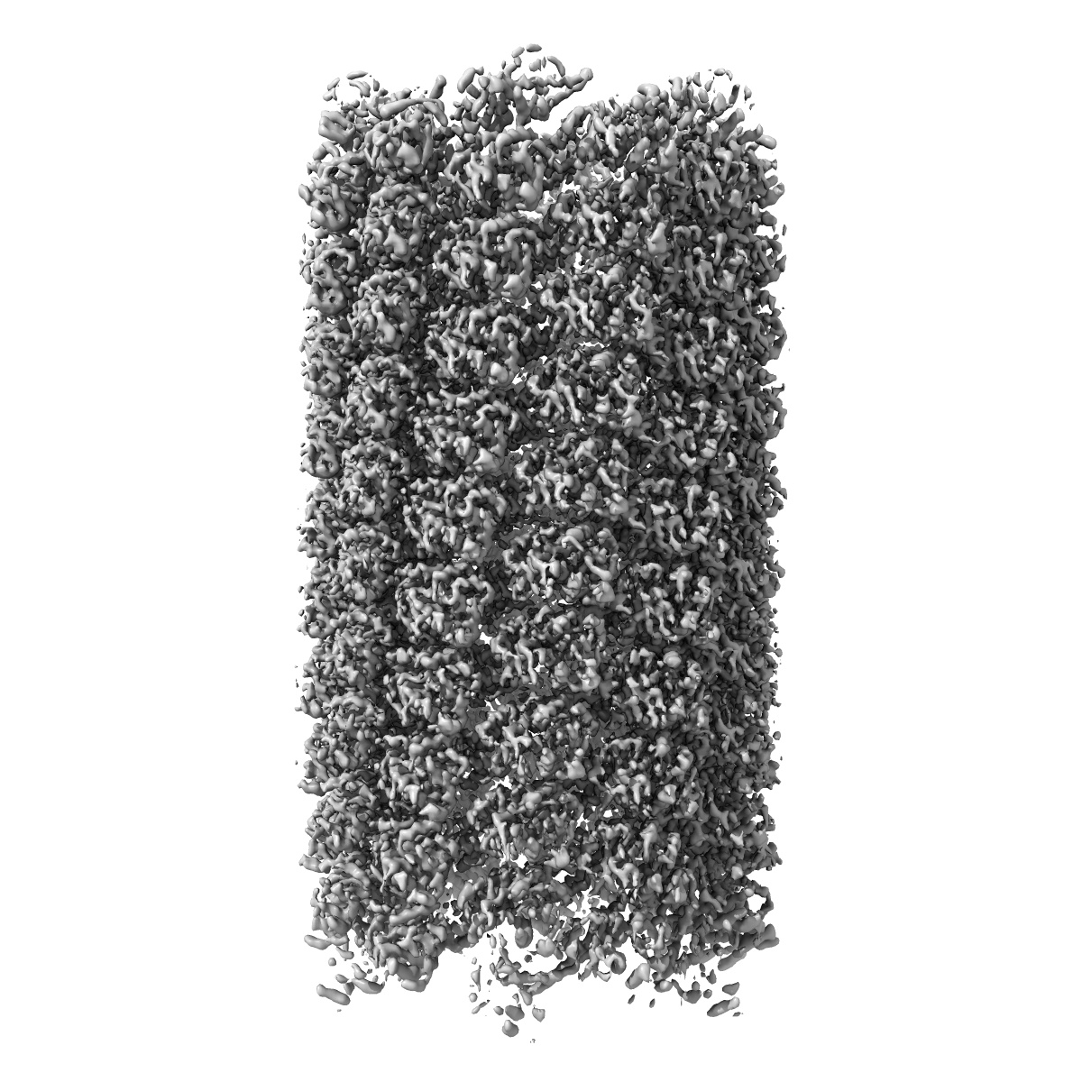
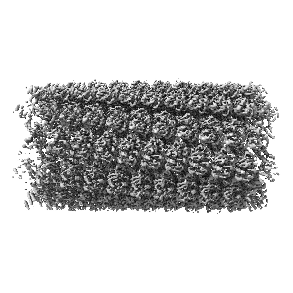
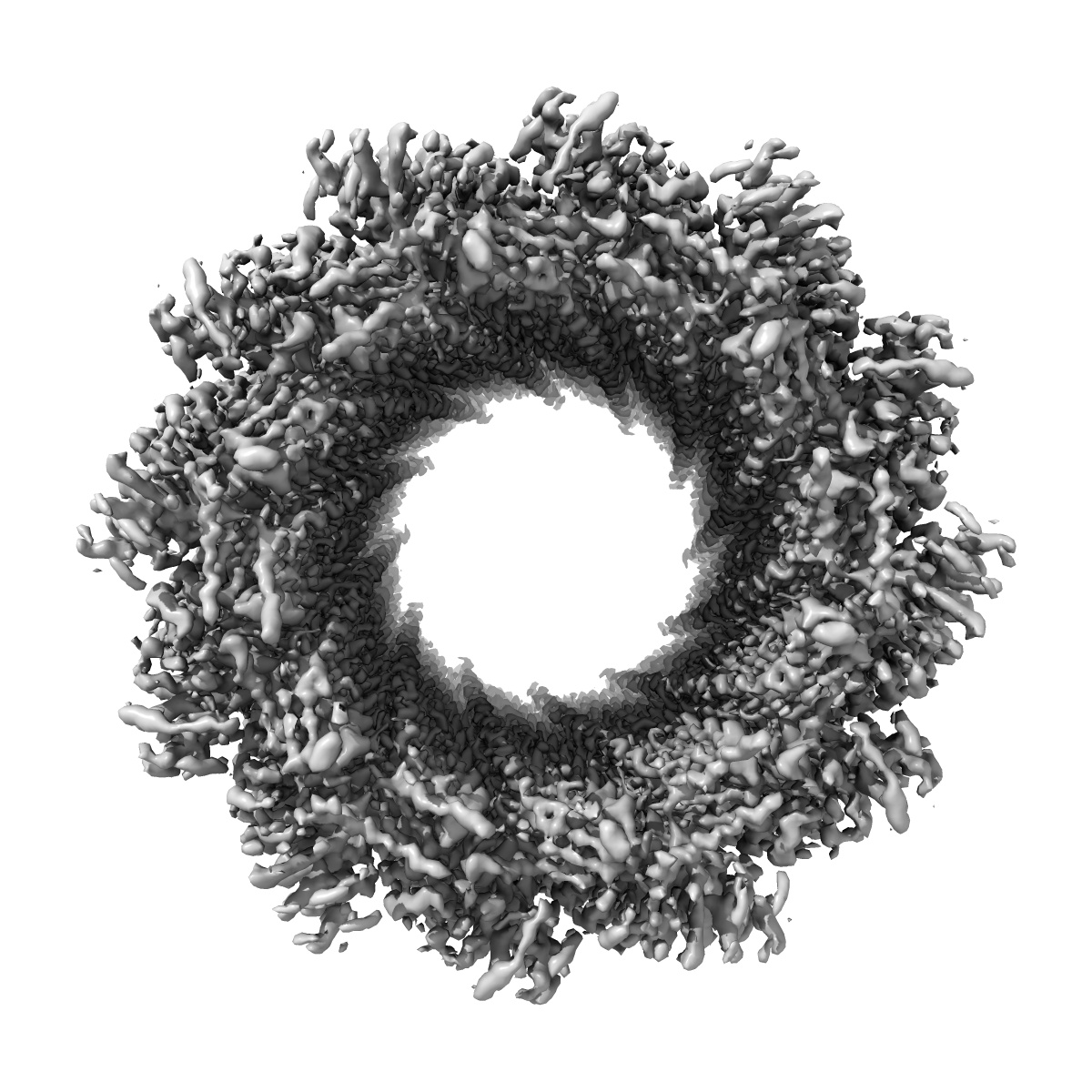
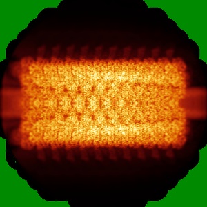
EMD-4859
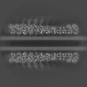
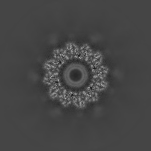
Cryo-EM structure of the anti-feeding prophage (AFP) sheath-tube in contracted state, C6 symmetrized
EMD-4859
Single-particle4.2 Å
 Deposition: 12/04/2019
Map released: 24/04/2019
Last modified: 06/11/2019
Buffer
pH: 7.0
Vitrification
Cryogen name: ETHANE
Microscope: FEI TITAN KRIOS
Illumination mode: FLOOD BEAM
Imaging mode: BRIGHT FIELD
Electron source: FIELD EMISSION GUN
Acceleration voltage: 300 kV
Illumination mode: FLOOD BEAM
Imaging mode: BRIGHT FIELD
Electron source: FIELD EMISSION GUN
Acceleration voltage: 300 kV
Image Recording
[1]
Final
reconstruction
Resolution: 4.2
Å
(
BY AUTHOR)
Resolution method: FSC 0.143 CUT-OFF
Number of images used: 15189
Resolution method: FSC 0.143 CUT-OFF
Number of images used: 15189
⌯ Applied Symmetry
Point group:
C6
Software
[1]
| Name | Version | Details |
|---|---|---|
| RELION | - | - |
Startup model
[1]
⦨ Initial angle
assignment
Type:
PROJECTION MATCHING
⦩ Final angle assignment
Type:
PROJECTION MATCHING
Particle selection
[1]
| Selected | Ref. model | Method | Software | Details |
|---|---|---|---|---|
| 15189 | - | - | - | - |
CTF correction
Software
[1]
| Name | Version | Details |
|---|---|---|
| CTFFIND | - | - |
Format: CCP4
Data type: IMAGE STORED AS FLOATING POINT NUMBER (4 BYTES)
Annotation details: None
Data type: IMAGE STORED AS FLOATING POINT NUMBER (4 BYTES)
Annotation details: None
⬡ Geometry
| X | Y | Z | |
|---|---|---|---|
| Dimensions | 364 | 364 | 364 |
| Origin | -182 | -182 | -182 |
| Spacing | 364 | 364 | 364 |
| Voxel size | 1.397 Å | 1.397 Å | 1.397 Å |
Contour list
| Primary | Level | Source |
|---|---|---|
| True | 0.12 | AUTHOR |