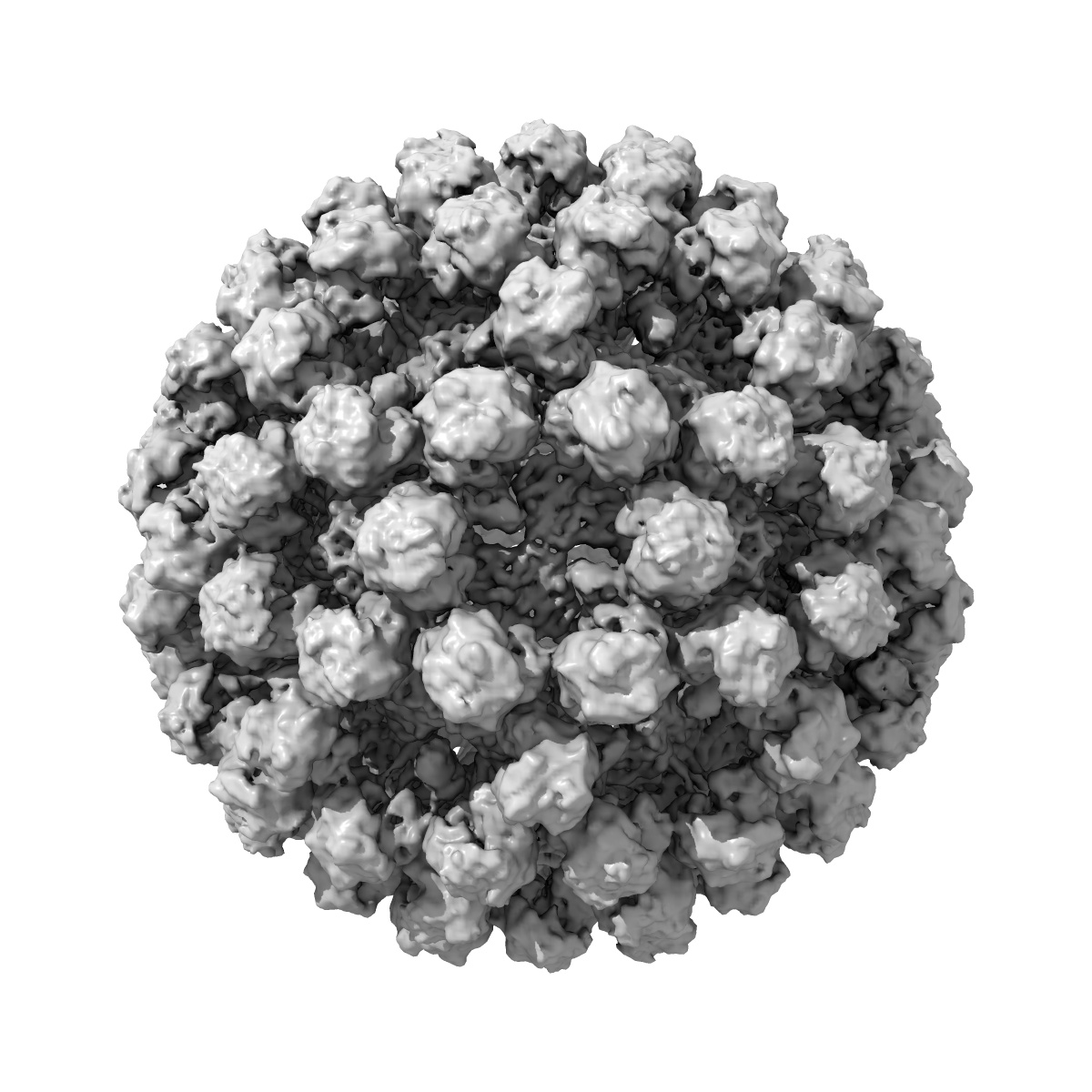
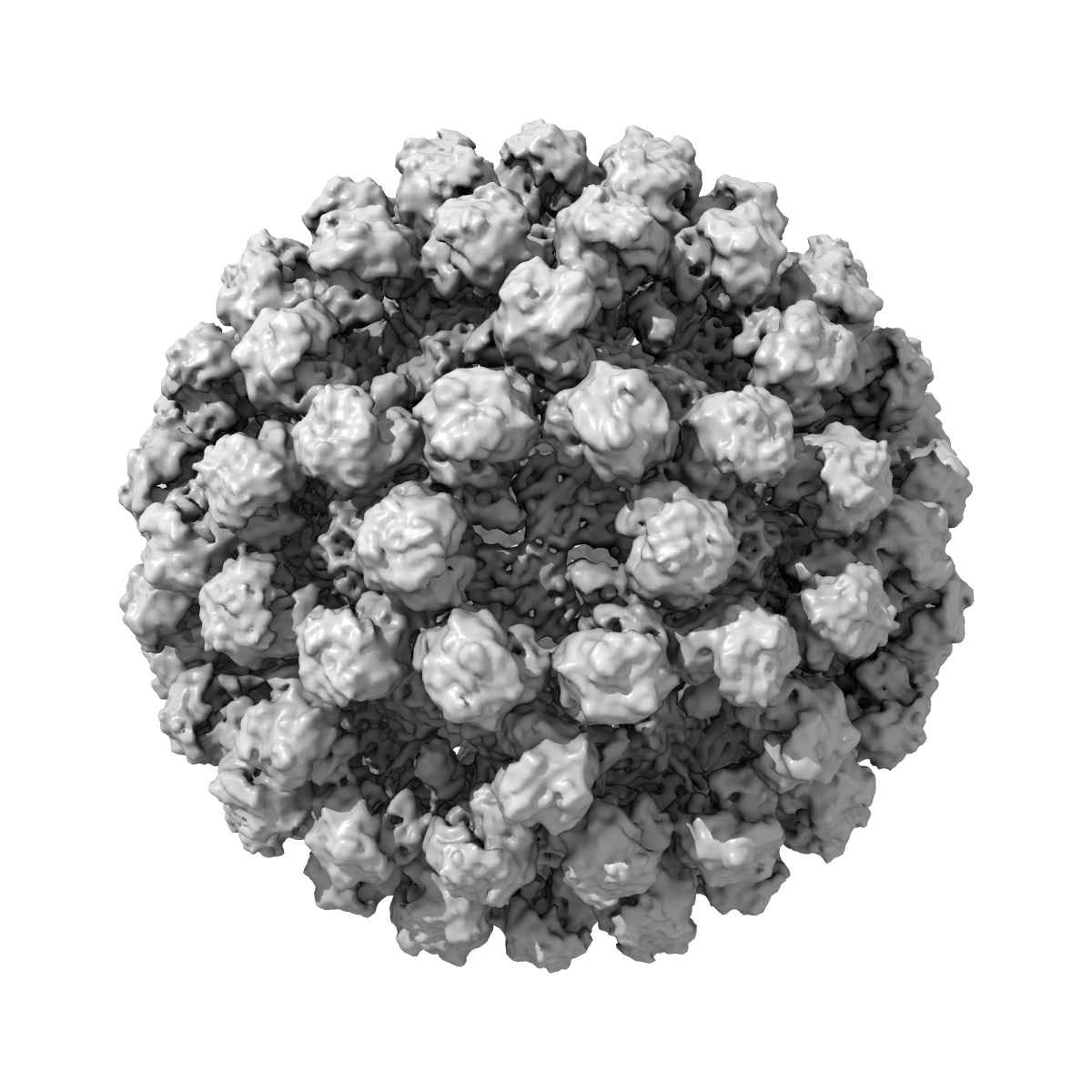
EMD-4550
Cryo-EM structure of human norovirus GII.4 NSW-2012 VLP with T=4 icosahedral symmetry
EMD-4550
Single-particle7.3 Å
 Deposition: 17/01/2019
Map released: 12/06/2019
Last modified: 26/06/2019
Concentration: 4
mg/mL
Buffer
pH: 7.4
Buffer components [1]:
Buffer components [1]:
| Name | Formula | Concentration | ChEBI |
|---|---|---|---|
| PBS | - | - | - |
Grid
Vitrification
Microscope: FEI TITAN KRIOS
Illumination mode: FLOOD BEAM
Imaging mode: BRIGHT FIELD
Electron source: FIELD EMISSION GUN
Acceleration voltage: 300 kV
Nominal CS: 2.7 mm
Nominal magnification: 64000.0
Illumination mode: FLOOD BEAM
Imaging mode: BRIGHT FIELD
Electron source: FIELD EMISSION GUN
Acceleration voltage: 300 kV
Nominal CS: 2.7 mm
Nominal magnification: 64000.0
Image Recording
[1]
Detector model:
GATAN K2 SUMMIT (4k x 4k)
Number of grids: 1
Number of real images: 364
Average electron dose per image: 20.0 e/Å2
Number of grids: 1
Number of real images: 364
Average electron dose per image: 20.0 e/Å2
Final
reconstruction
Resolution: 7.3
Å
(
BY AUTHOR)
Resolution method: FSC 0.143 CUT-OFF
Number of images used: 10548
Resolution method: FSC 0.143 CUT-OFF
Number of images used: 10548
⌯ Applied Symmetry
Point group:
I
Software
[1]
| Name | Version | Details |
|---|---|---|
| RELION | 2.1 | - |
⦨ Initial angle
assignment
Type:
MAXIMUM LIKELIHOOD
⦩ Final angle assignment
Type:
MAXIMUM LIKELIHOOD
Format: CCP4
Data type: IMAGE STORED AS FLOATING POINT NUMBER (4 BYTES)
Annotation details: Cryo-EM structure of GII.4 NSW virus like particle
Data type: IMAGE STORED AS FLOATING POINT NUMBER (4 BYTES)
Annotation details: Cryo-EM structure of GII.4 NSW virus like particle
⬡ Geometry
| X | Y | Z | |
|---|---|---|---|
| Dimensions | 256 | 256 | 256 |
| Origin | 0 | 0 | 0 |
| Spacing | 256 | 256 | 256 |
| Voxel size | 2.27 Å | 2.27 Å | 2.27 Å |
Contour list
| Primary | Level | Source |
|---|---|---|
| True | 0.05 | AUTHOR |