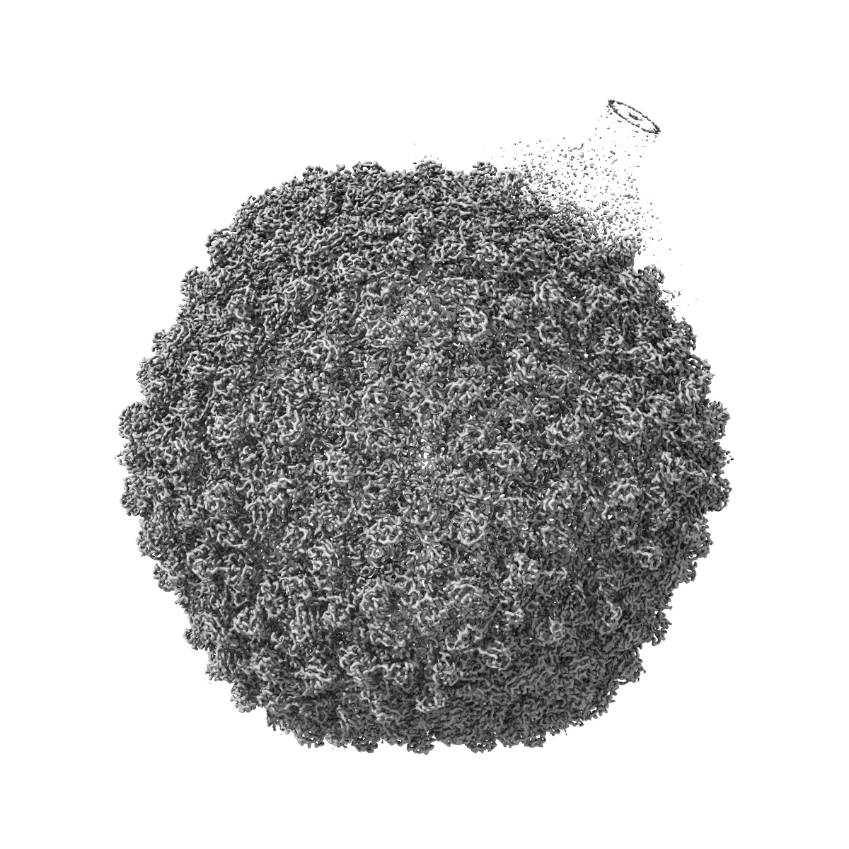
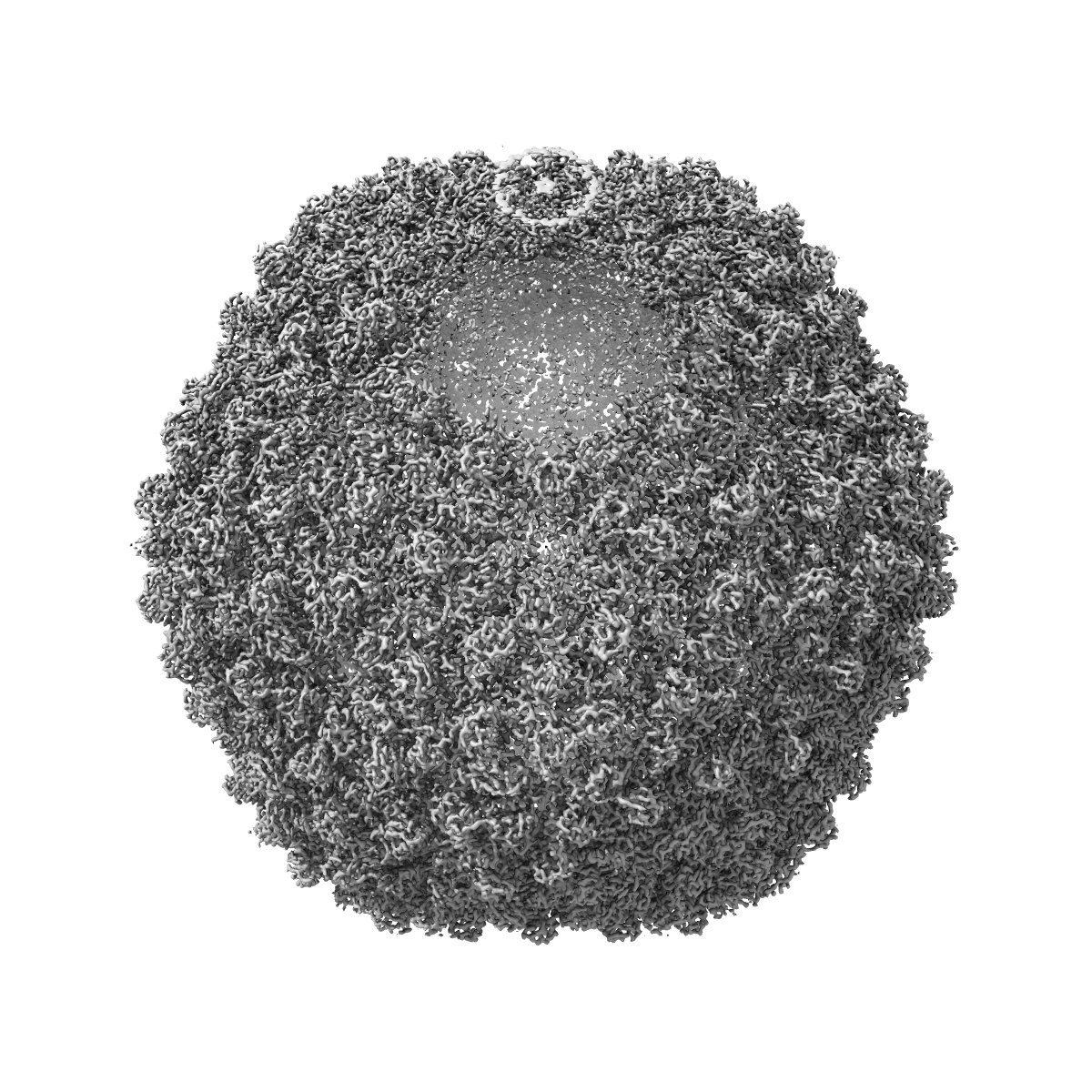
EMD-4436
Five-fold symmetrized map of native bacteriophage P68
EMD-4436
Single-particle3.8 Å
 Deposition: 26/11/2018
Map released: 06/11/2019
Last modified: 13/11/2019
Concentration: 2
mg/mL
Buffer
pH: 8.0
Buffer components [3]:
Buffer components [3]:
| Name | Formula | Concentration | ChEBI |
|---|---|---|---|
| Tris(hydroxymethyl)aminomethane | Tris | 50.0 mM | |
| Calcium chloride | CaCl | 10.0 mM | |
| Sodium chloride | NaCl | 10.0 mM |
Vitrification
Cryogen name: ETHANE
Chamber humidity: 100%
Chamber temperature: 293 K
Instrument: FEI VITROBOT MARK IV
Details: Blot time 2s; blot force -2; 3.6 ul of sample.
Chamber humidity: 100%
Chamber temperature: 293 K
Instrument: FEI VITROBOT MARK IV
Details: Blot time 2s; blot force -2; 3.6 ul of sample.
Microscope: FEI TITAN KRIOS
Illumination mode: FLOOD BEAM
Imaging mode: BRIGHT FIELD
Electron source: FIELD EMISSION GUN
Acceleration voltage: 300 kV
C2 aperture diameter: 70.0 µm
Nominal CS: 2.7 mm
Nominal defocus: 0.001 µm - 0.003 µm
Nominal magnification: 75000.0
Specimen holder model: FEI TITAN KRIOS AUTOGRID HOLDER
Cooling holder cryogen: NITROGEN
Alignment procedure: COMA FREE
Image Recording
[1]
Detector model:
FEI FALCON II (4k x 4k)
Detector mode: INTEGRATING
Dimensions: 4096 pixel x 4096 pixel
Sampling interval: 1.063 µm
Frames per image: 1-7
Number of grids: 2
Number of real images: 2891
Average exposure time: 1.0 s
Average electron dose per image: 21.0 e/Å2
Detector mode: INTEGRATING
Dimensions: 4096 pixel x 4096 pixel
Sampling interval: 1.063 µm
Frames per image: 1-7
Number of grids: 2
Number of real images: 2891
Average exposure time: 1.0 s
Average electron dose per image: 21.0 e/Å2
Final
reconstruction
Resolution: 3.8
Å
(
BY AUTHOR)
Resolution method: FSC 0.143 CUT-OFF
Number of classed used: 1
Number of images used: 28826
Algorithm: BACK PROJECTION
Resolution method: FSC 0.143 CUT-OFF
Number of classed used: 1
Number of images used: 28826
Algorithm: BACK PROJECTION
⌯ Applied Symmetry
Point group:
C5
Software
[1]
| Name | Version | Details |
|---|---|---|
| RELION | 2.1 | - |
Startup model
[1]
⦨ Initial angle
assignment
⦩ Final angle assignment
Particle selection
[1]
| Selected | Ref. model | Method | Software | Details |
|---|---|---|---|---|
| 37218 | - | - | - | - |
Final 3D classification
Number of classes:
3
Avg. number of members per classes: 11210.0
Avg. number of members per classes: 11210.0
Software
[1]
| Name | Version | Details |
|---|---|---|
| RELION | 2.1 | - |
CTF correction
Software
[1]
| Name | Version | Details |
|---|---|---|
| CTFFIND | 4.0 | - |
Format: CCP4
Data type: IMAGE STORED AS FLOATING POINT NUMBER (4 BYTES)
Annotation details: Five-fold symmetrized map of native bacteriophage P68
Data type: IMAGE STORED AS FLOATING POINT NUMBER (4 BYTES)
Annotation details: Five-fold symmetrized map of native bacteriophage P68
⬡ Geometry
| X | Y | Z | |
|---|---|---|---|
| Dimensions | 600 | 600 | 600 |
| Origin | -300 | -300 | -300 |
| Spacing | 600 | 600 | 600 |
| Voxel size | 1.063 Å | 1.063 Å | 1.063 Å |
Contour list
| Primary | Level | Source |
|---|---|---|
| True | 0.1 | AUTHOR |