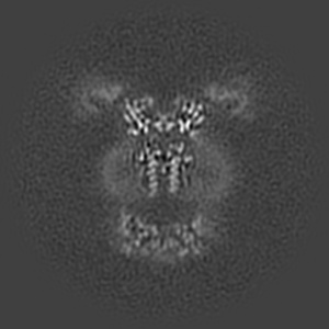
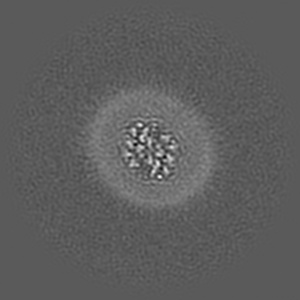
EMD-4246
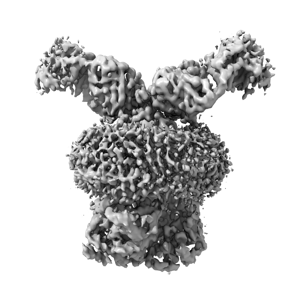
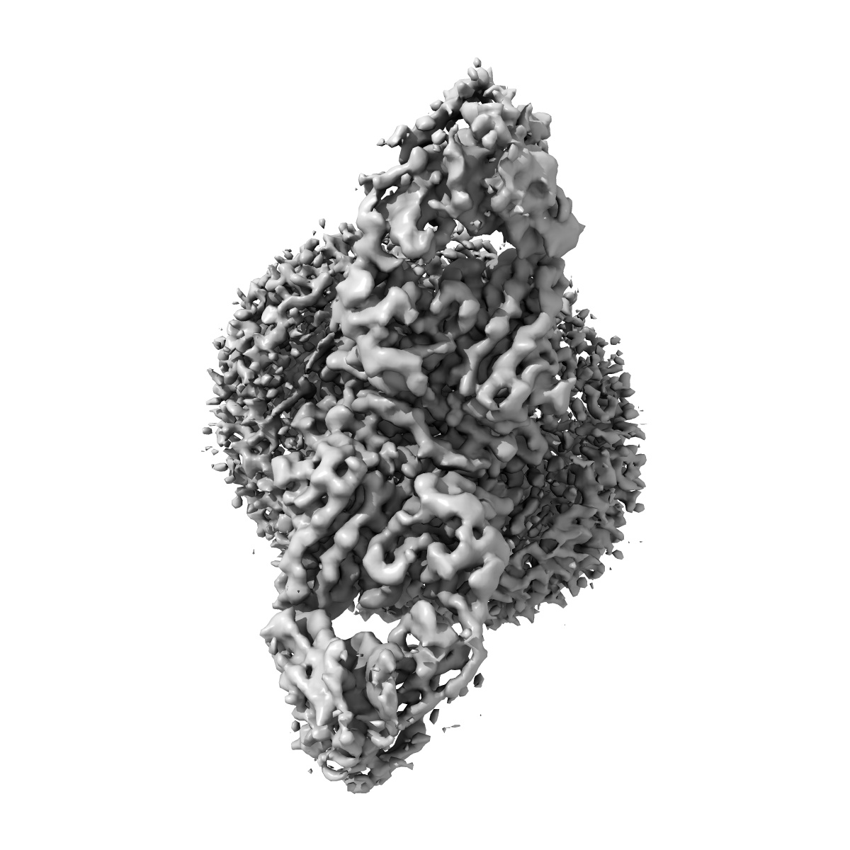
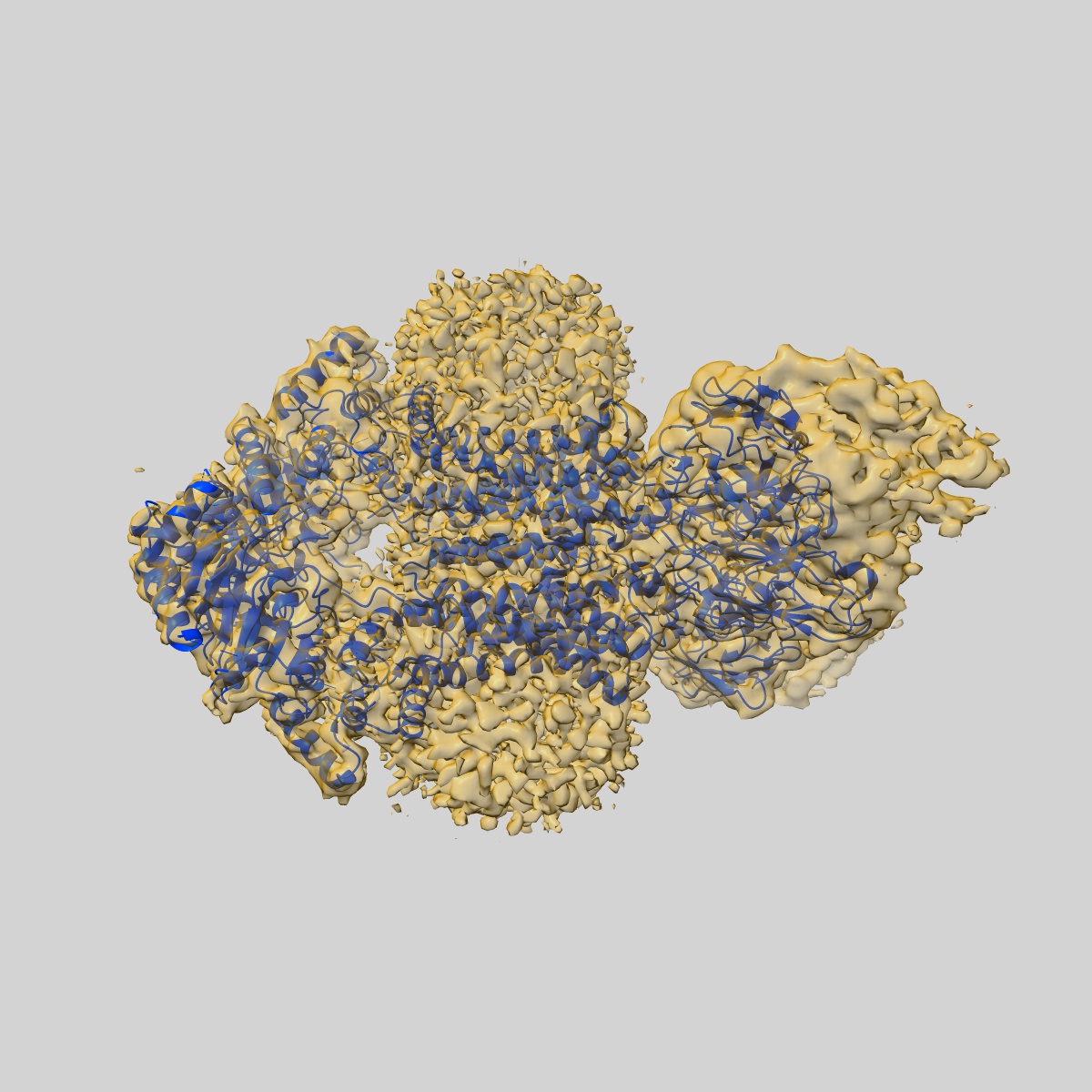
Structure of inhibitor-bound ABCG2
EMD-4246
Single-particle3.6 Å
 Deposition: 03/01/2018
Map released: 11/04/2018
Last modified: 29/07/2020
Name: inhibitor-bound ABCG2
Summary:
Name: inhibitor-bound ABCG2
Name: ATP-binding cassette sub-family G member 2
Number of copies: 2
UniProtKB PDBe-KB AlphaFold DB
Number of copies: 2
UniProtKB PDBe-KB AlphaFold DB
Domains
Gene Ontology [34]
Biological process:
transmembrane transport biotin transport urate metabolic process transport across blood-brain barrier xenobiotic transport across blood-brain barrier lipid transport export across plasma membrane transepithelial transport riboflavin transport cellular detoxification organic anion transport urate salt excretion renal urate salt excretion
Molecular function:
ATP hydrolysis activity protein homodimerization activity efflux transmembrane transporter activity ABC-type xenobiotic transporter activity organic anion transmembrane transporter activity urate transmembrane transporter activity riboflavin transmembrane transporter activity ATP binding biotin transmembrane transporter activity xenobiotic transmembrane transporter activity identical protein binding ATPase-coupled transmembrane transporter activity
Cellular location:
membrane external side of apical plasma membrane membrane raft plasma membrane nucleoplasm mitochondrial membrane integral component of membrane brush border membrane apical plasma membrane
Natural source
Organism: Homo sapiens
Molecular weight
| Experimental | Theoretical | Method |
|---|---|---|
| - | 72 kDa | - |
Recombinant Expression
| Organism | Strain | Cell | Plasmid |
|---|---|---|---|
| Homo sapiens | - | - | - |
Name: 5D3(Fab) light chain variable domain
Number of copies: 2
Details: The variable domain of the light chain of 5D3(Fab)
Number of copies: 2
Details: The variable domain of the light chain of 5D3(Fab)
Natural source
Organism: Mus musculus
Molecular weight
| Experimental | Theoretical | Method |
|---|---|---|
| - | 23 kDa | - |
Recombinant Expression
| Organism | Strain | Cell | Plasmid |
|---|---|---|---|
| Mus musculus | - | - | - |
Name: 5D3(Fab) heavy chain variable domain
Number of copies: 2
Details: The variable domain of the heavy chain of 5D3(Fab)
UniProtKB PDBe-KB AlphaFold DB
Number of copies: 2
Details: The variable domain of the heavy chain of 5D3(Fab)
UniProtKB PDBe-KB AlphaFold DB
Natural source
Organism: Mus musculus
Molecular weight
| Experimental | Theoretical | Method |
|---|---|---|
| - | 23 kDa | - |
Recombinant Expression
| Organism | Strain | Cell | Plasmid |
|---|---|---|---|
| Mus musculus | - | - | - |
Name: 2-acetamido-2-deoxy-beta-D-glucopyranose
HET code: NAG
Number of copies: 2
ChEMBL ChEBI
HET code: NAG
Number of copies: 2
ChEMBL ChEBI
Molecular weight
| Experimental | Theoretical | Method |
|---|---|---|
| - | 221 Da | - |
Name: ~{N}-[5-[1-[4-[2-[6-methoxy-7-[2-[2-(2-methoxyethoxy)ethoxy]ethoxy]-3,4-dihydro-1~{H}-isoquinolin-2-yl]ethyl]phenyl]-1,2,3-triazol-4-yl]-2-propanoyl-phenyl]quinoline-2-carboxamide
HET code: D6T
Number of copies: 1
ChEMBL
HET code: D6T
Number of copies: 1
ChEMBL
Molecular weight
| Experimental | Theoretical | Method |
|---|---|---|
| - | 798 Da | - |