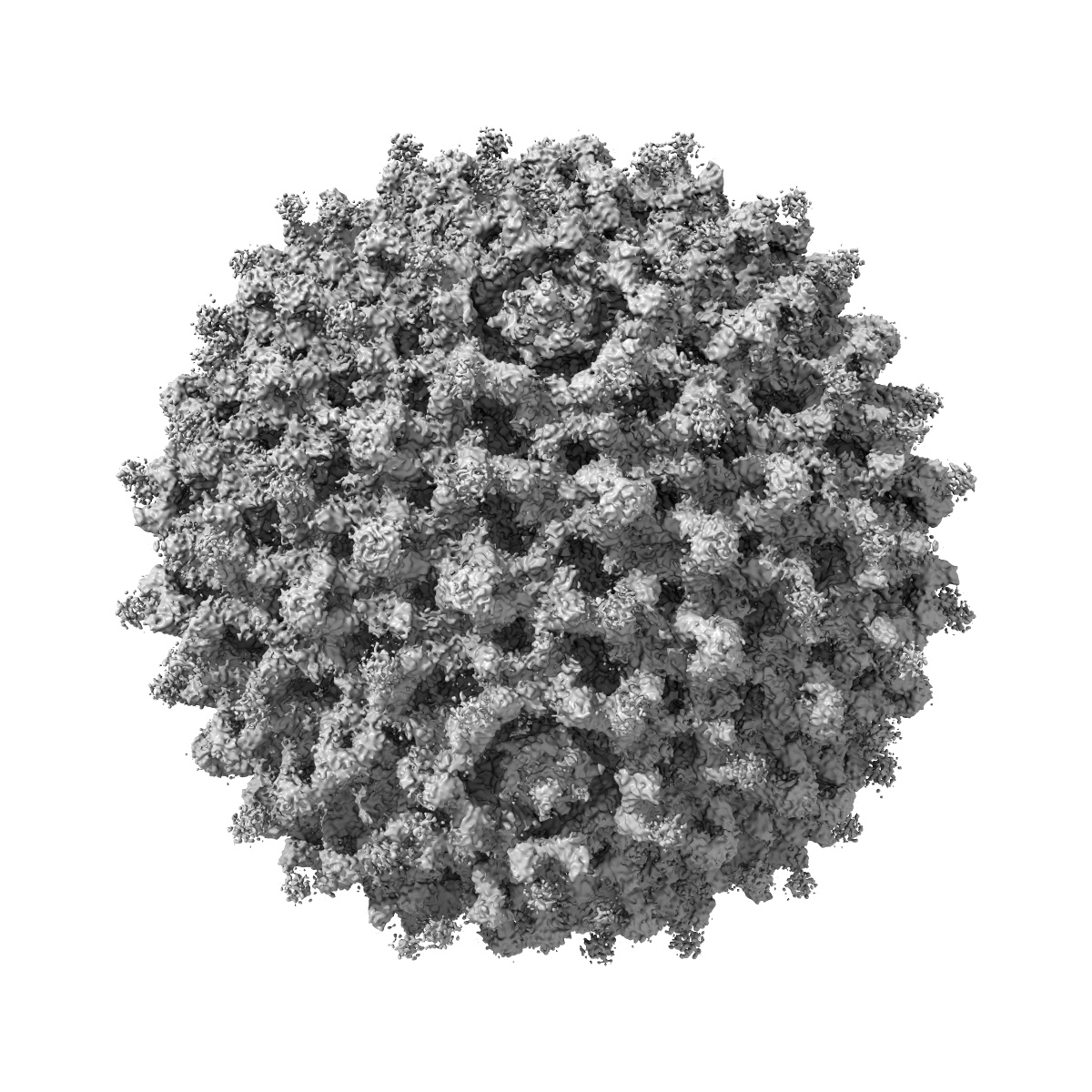
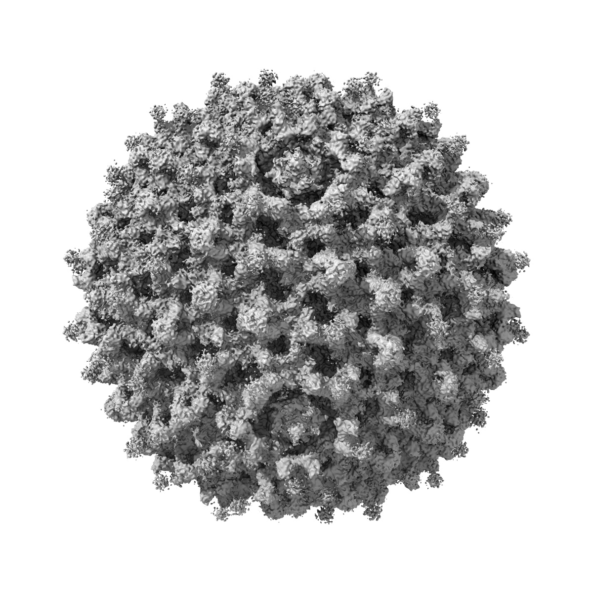
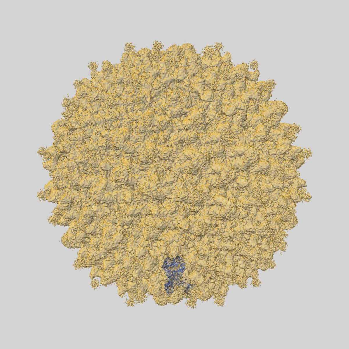
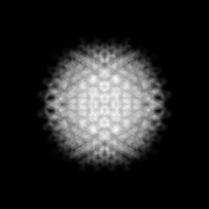
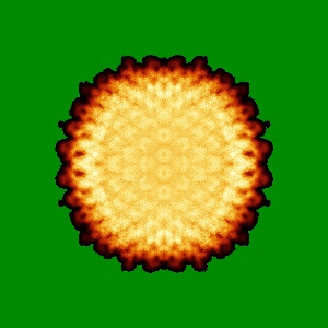
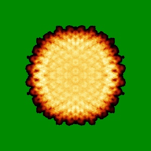
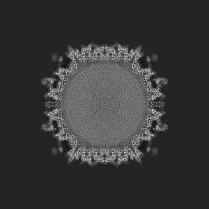
EMD-3821
Cryo-EM reconstruction of a modified human Adenovirus C5
EMD-3821
Single-particle7.4 Å
 Deposition: 21/07/2017
Map released: 07/02/2018
Last modified: 25/11/2020
Buffer
pH: 7.4
Vitrification
Cryogen name: ETHANE
Microscope: FEI TITAN KRIOS
Illumination mode: FLOOD BEAM
Imaging mode: BRIGHT FIELD
Electron source: FIELD EMISSION GUN
Acceleration voltage: 300 kV
Illumination mode: FLOOD BEAM
Imaging mode: BRIGHT FIELD
Electron source: FIELD EMISSION GUN
Acceleration voltage: 300 kV
Image Recording
[1]
Detector model:
GATAN K2 SUMMIT (4k x 4k)
Detector mode: SUPER-RESOLUTION
Average exposure time: 0.4 s
Average electron dose per image: 1.0 e/Å2
Detector mode: SUPER-RESOLUTION
Average exposure time: 0.4 s
Average electron dose per image: 1.0 e/Å2
Final
reconstruction
Resolution: 7.4
Å
(
BY AUTHOR)
Resolution method: FSC 0.143 CUT-OFF
Number of images used: 1880
Resolution method: FSC 0.143 CUT-OFF
Number of images used: 1880
Software
[1]
| Name | Version | Details |
|---|---|---|
| RELION | 1.4 | - |
⦨ Initial angle
assignment
Type:
ANGULAR RECONSTITUTION
⦩ Final angle assignment
Type:
ANGULAR RECONSTITUTION
CTF correction
Software
[1]
| Name | Version | Details |
|---|---|---|
| CTFFIND | 4.0.17 | - |
Format: CCP4
Data type: IMAGE STORED AS FLOATING POINT NUMBER (4 BYTES)
Data type: IMAGE STORED AS FLOATING POINT NUMBER (4 BYTES)
⬡ Geometry
| X | Y | Z | |
|---|---|---|---|
| Dimensions | 800 | 800 | 800 |
| Origin | 0 | 0 | 0 |
| Spacing | 800 | 800 | 800 |
| Voxel size | 1.96 Å | 1.96 Å | 1.96 Å |
Contour list
| Primary | Level | Source |
|---|---|---|
| True | 0.014 | AUTHOR |