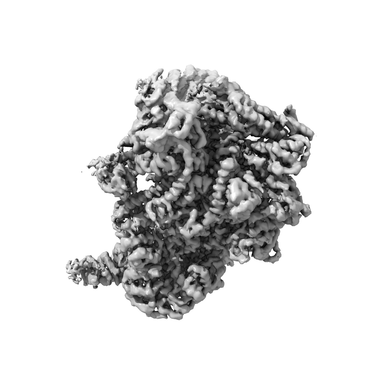
EMD-3388
Cryo-EM structure of combined apo phosphorylated APC/C
EMD-3388
Single-particle3.4 Å
 Deposition: 15/03/2016
Map released: 04/05/2016
Last modified: 01/06/2016
Concentration: 0.15
mg/mL
Buffer
pH: 8.0
Details: 20mM Hepes, 150mM NaCl, 0.005mM TCEP
Details: 20mM Hepes, 150mM NaCl, 0.005mM TCEP
Staining
Type:
NEGATIVE
Details: Vitrification in liquid ethane
Details: Vitrification in liquid ethane
Grid
Details: 200 mesh Quantifoil R2/2 copper grid with thin carbon support, treated with a 9:1 argon:oxygen plasma cleaner for 20-40s
Vitrification
Cryogen name: ETHANE
Chamber humidity: 100%
Chamber temperature: 100 K
Instrument: FEI VITROBOT MARK III
Method: The grids were incubated for 30s at 4 degree and 100% humidity before blotting for 5s and plunging into liquid ethane
Chamber humidity: 100%
Chamber temperature: 100 K
Instrument: FEI VITROBOT MARK III
Method: The grids were incubated for 30s at 4 degree and 100% humidity before blotting for 5s and plunging into liquid ethane
Microscope: FEI POLARA 300
Illumination mode: FLOOD BEAM
Imaging mode: BRIGHT FIELD
Electron source: FIELD EMISSION GUN
Acceleration voltage: 300 kV
Nominal CS: 2 mm
Nominal defocus: 2.0 µm - 4.0 µm
Nominal magnification: 78000.0
Specimen holder model: OTHER
Specimen holder details: Liquid nitrogen cooled
Illumination mode: FLOOD BEAM
Imaging mode: BRIGHT FIELD
Electron source: FIELD EMISSION GUN
Acceleration voltage: 300 kV
Nominal CS: 2 mm
Nominal defocus: 2.0 µm - 4.0 µm
Nominal magnification: 78000.0
Specimen holder model: OTHER
Specimen holder details: Liquid nitrogen cooled
Temperature
Minimum: 90
K
Average: 105 K
Maximum: 120 K
Average: 105 K
Maximum: 120 K
Specialist optics
Energy filter
Name:
FEI
Image Recording
[1]
Detector category:
CCD
Detector model: FEI FALCON II (4k x 4k)
Number of real images: 12500
Average electron dose per image: 27 e/Å2
Details: The exposure time for each micrograph was 2s at a dose rate of 27 electrons/angstrom2/s. 34 movie frames were recorded for each micrograph.
Detector model: FEI FALCON II (4k x 4k)
Number of real images: 12500
Average electron dose per image: 27 e/Å2
Details: The exposure time for each micrograph was 2s at a dose rate of 27 electrons/angstrom2/s. 34 movie frames were recorded for each micrograph.
Details: Particles were selected by automatic particle picking in RELION
Final
reconstruction
CTF correction
Details:Each micrograph
Format: CCP4
Data type: IMAGE STORED AS FLOATING POINT NUMBER (4 BYTES)
Annotation details: Reconstruction of apo phosphorylated APC/C using both apo phosphorylated APC/C data and apo phosphorylated particles classified from APC/C-Cdc20-Hsl1
Details: ::::EMDATABANK.org::::EMD-3388::::
Data type: IMAGE STORED AS FLOATING POINT NUMBER (4 BYTES)
Annotation details: Reconstruction of apo phosphorylated APC/C using both apo phosphorylated APC/C data and apo phosphorylated particles classified from APC/C-Cdc20-Hsl1
Details: ::::EMDATABANK.org::::EMD-3388::::
⬡ Geometry
| X | Y | Z | |
|---|---|---|---|
| Dimensions | 284 | 284 | 284 |
| Origin | 0 | 0 | 0 |
| Spacing | 284 | 284 | 284 |
| Voxel size | 1.36 Å | 1.36 Å | 1.36 Å |
Contour list
| Primary | Level | Source |
|---|---|---|
| True | 0.08 | AUTHOR |