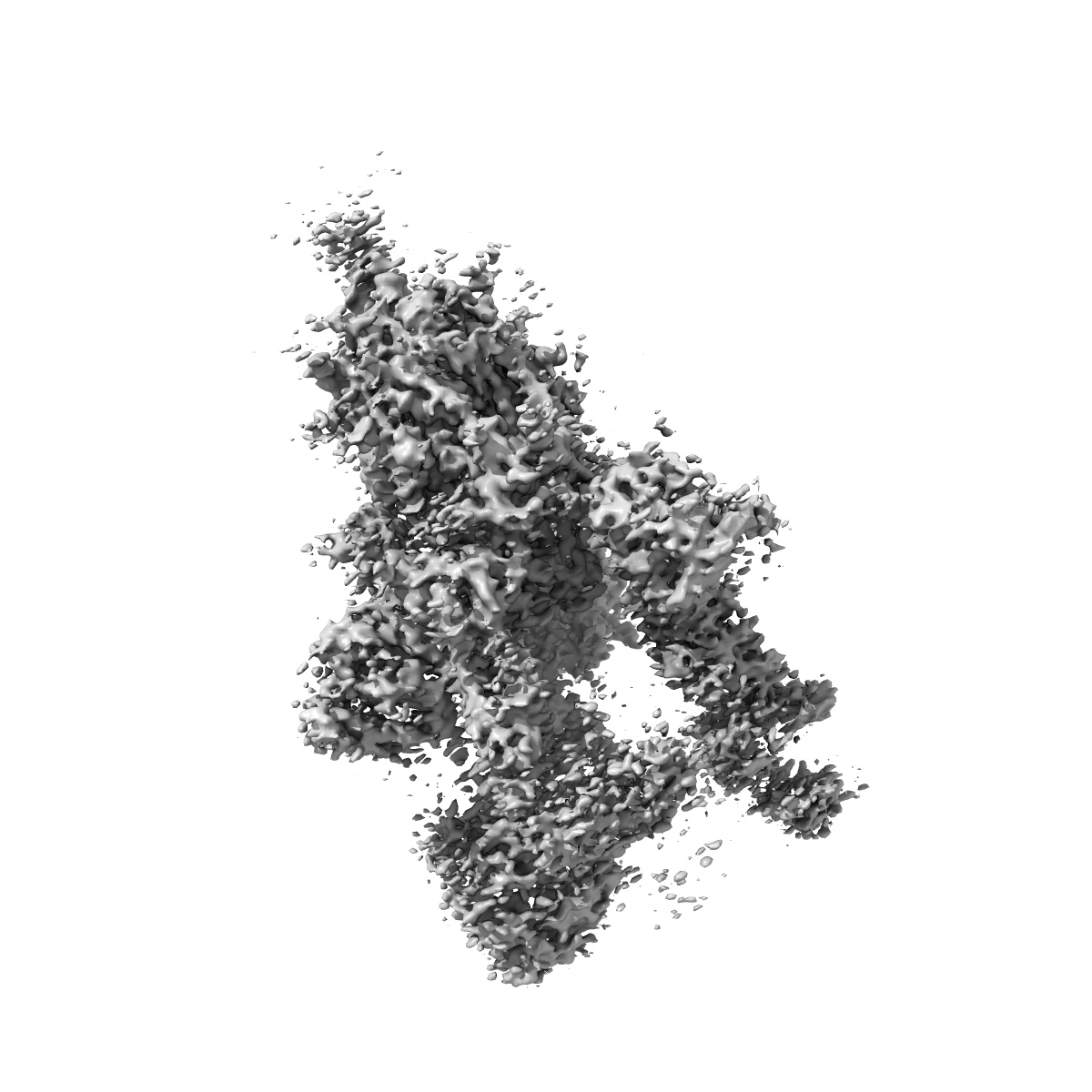
EMD-30460
cryo EM map of the S protein of SARS-CoV-2 in complex bound with T-ACE2
EMD-30460
Single-particle4.0 Å
 Deposition: 18/08/2020
Map released: 18/11/2020
Last modified: 10/02/2021
Buffer
pH: 8.0
Vitrification
Cryogen name: ETHANE
Microscope: FEI TITAN KRIOS
Illumination mode: FLOOD BEAM
Imaging mode: BRIGHT FIELD
Electron source: FIELD EMISSION GUN
Acceleration voltage: 300 kV
Specimen holder model: FEI TITAN KRIOS AUTOGRID HOLDER
Cooling holder cryogen: NITROGEN
Alignment procedure: COMA FREE
Illumination mode: FLOOD BEAM
Imaging mode: BRIGHT FIELD
Electron source: FIELD EMISSION GUN
Acceleration voltage: 300 kV
Specimen holder model: FEI TITAN KRIOS AUTOGRID HOLDER
Cooling holder cryogen: NITROGEN
Alignment procedure: COMA FREE
Image Recording
[1]
Final
reconstruction
Resolution: 4.0
Å
(
BY AUTHOR)
Resolution method: FSC 0.143 CUT-OFF
Number of images used: 57404
Resolution method: FSC 0.143 CUT-OFF
Number of images used: 57404
Software
[1]
| Name | Version | Details |
|---|---|---|
| RELION | 3.0.6 | - |
⦨ Initial angle
assignment
Type:
ANGULAR RECONSTITUTION
⦩ Final angle assignment
Type:
MAXIMUM LIKELIHOOD
Format: CCP4
Data type: IMAGE STORED AS FLOATING POINT NUMBER (4 BYTES)
Annotation details: Cryo EM map of the S protein of SARS-CoV-2 in complex bound with T-ACE2
Data type: IMAGE STORED AS FLOATING POINT NUMBER (4 BYTES)
Annotation details: Cryo EM map of the S protein of SARS-CoV-2 in complex bound with T-ACE2
⬡ Geometry
| X | Y | Z | |
|---|---|---|---|
| Dimensions | 288 | 288 | 288 |
| Origin | 0 | 0 | 0 |
| Spacing | 288 | 288 | 288 |
| Voxel size | 1.087 Å | 1.087 Å | 1.087 Å |
Contour list
| Primary | Level | Source |
|---|---|---|
| True | 0.015 | AUTHOR |