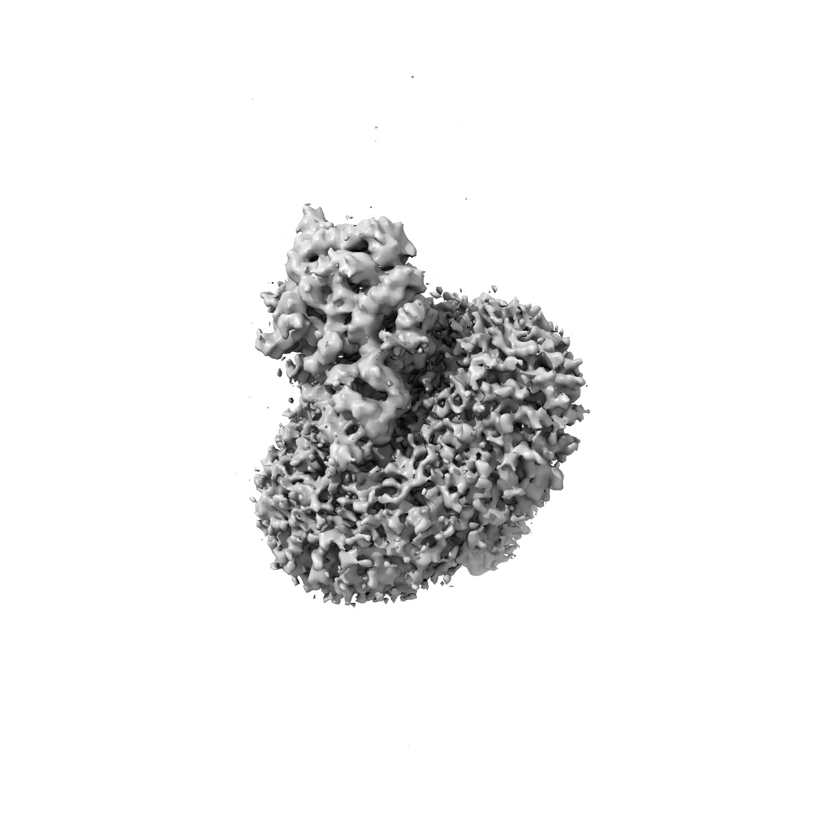
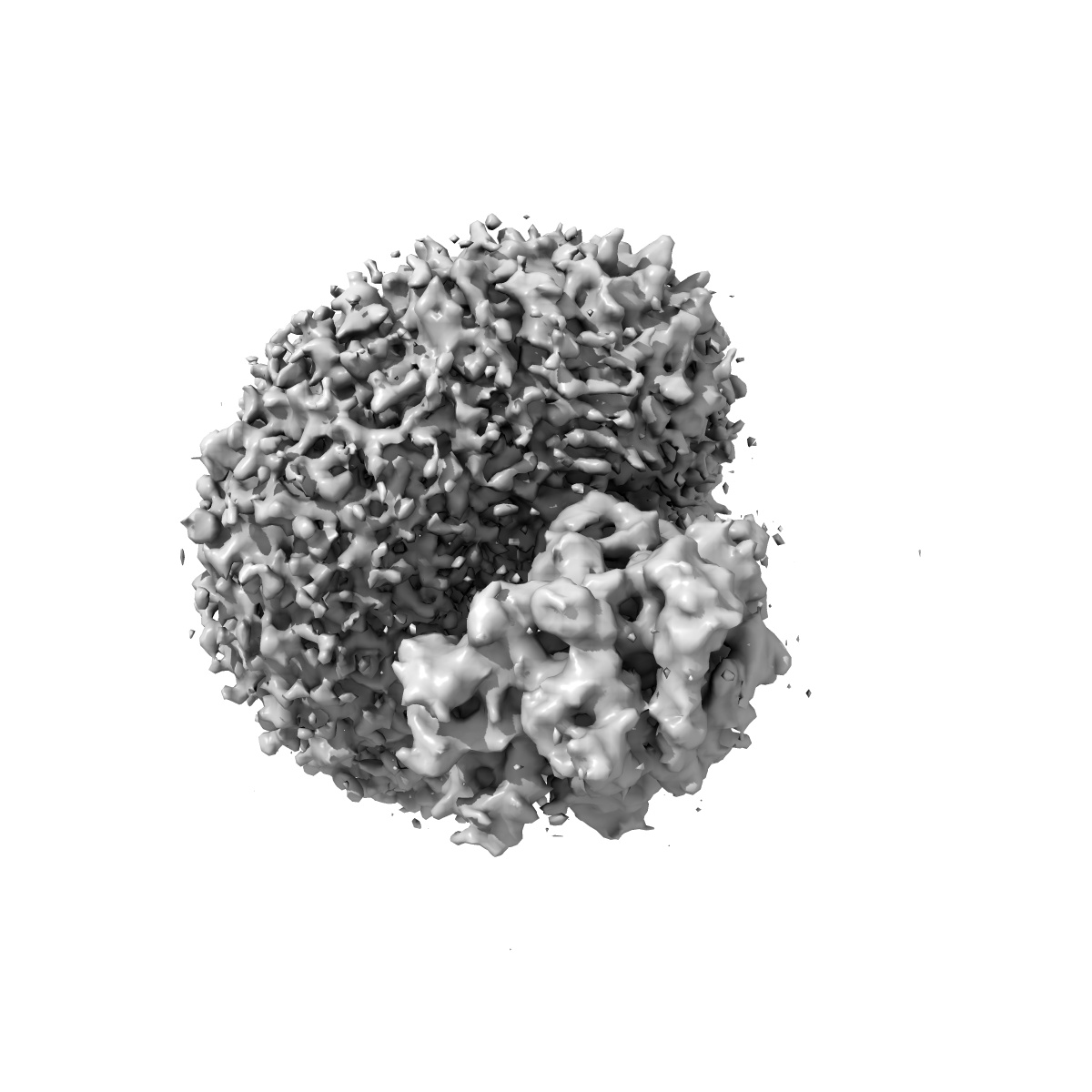
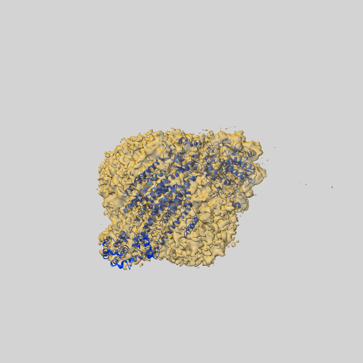
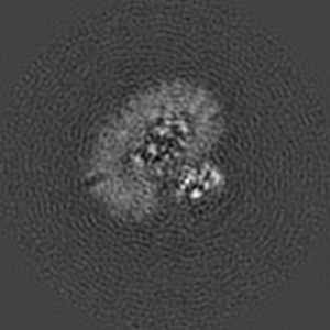
EMD-2974
The cryoEM map of human gamma-Secretase complex
EMD-2974
Single-particle4.4 Å
 Deposition: 03/04/2015
Map released: 17/06/2015
Last modified: 12/08/2015
Concentration: 4.2
mg/mL
Buffer
pH: 7.4
Details: 0.1% digitonin, 25 mM HEPES, pH 7.4, and 150 mM NaCl.
Details: 0.1% digitonin, 25 mM HEPES, pH 7.4, and 150 mM NaCl.
Grid
Details: Quantifoil Cu R1.2/1.3 grids
Vitrification
Cryogen name: ETHANE
Chamber humidity: 100%
Chamber temperature: 277 K
Instrument: FEI VITROBOT MARK IV
Method: Blot for 3 seconds before plunging
Chamber humidity: 100%
Chamber temperature: 277 K
Instrument: FEI VITROBOT MARK IV
Method: Blot for 3 seconds before plunging
Microscope: FEI TITAN KRIOS
Illumination mode: SPOT SCAN
Imaging mode: BRIGHT FIELD
Electron source: FIELD EMISSION GUN
Acceleration voltage: 300 kV
Nominal CS: 1.4 mm
Nominal defocus: 1.5 µm - 3.0 µm
Specimen holder model: FEI TITAN KRIOS AUTOGRID HOLDER
Illumination mode: SPOT SCAN
Imaging mode: BRIGHT FIELD
Electron source: FIELD EMISSION GUN
Acceleration voltage: 300 kV
Nominal CS: 1.4 mm
Nominal defocus: 1.5 µm - 3.0 µm
Specimen holder model: FEI TITAN KRIOS AUTOGRID HOLDER
Image Recording
[1]
Detector category:
CCD
Detector model: DIRECT ELECTRON DE-12 (4k x 3k)
Number of real images: 3312
Average electron dose per image: 4.5 e/Å2
Detector model: DIRECT ELECTRON DE-12 (4k x 3k)
Number of real images: 3312
Average electron dose per image: 4.5 e/Å2
Format: CCP4
Data type: IMAGE STORED AS FLOATING POINT NUMBER (4 BYTES)
Annotation details: Reconstruction of T4-lysozyme fusion gamma-secretase
Details: ::::EMDATABANK.org::::EMD-2974::::
Data type: IMAGE STORED AS FLOATING POINT NUMBER (4 BYTES)
Annotation details: Reconstruction of T4-lysozyme fusion gamma-secretase
Details: ::::EMDATABANK.org::::EMD-2974::::
⬡ Geometry
| X | Y | Z | |
|---|---|---|---|
| Dimensions | 200 | 200 | 200 |
| Origin | 0 | 0 | 0 |
| Spacing | 200 | 200 | 200 |
| Voxel size | 1.32 Å | 1.32 Å | 1.32 Å |
Contour list
| Primary | Level | Source |
|---|---|---|
| True | 0.02 | AUTHOR |