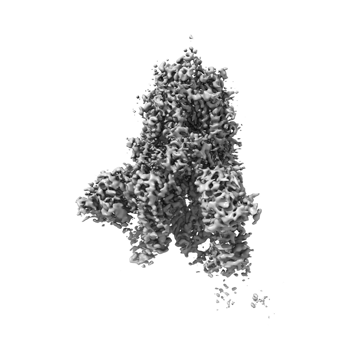
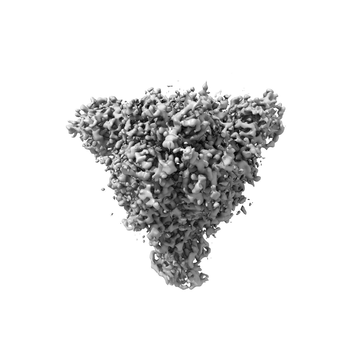
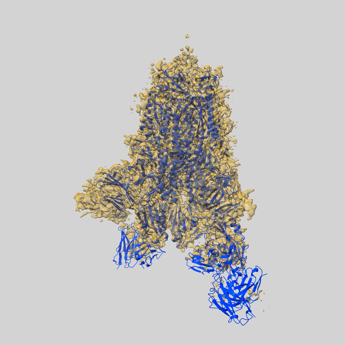
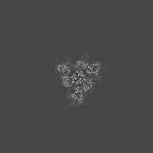
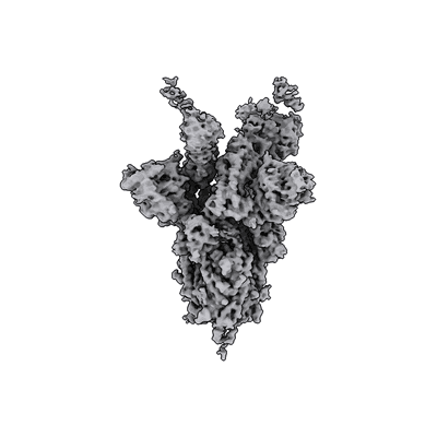
EMD-22492
SARS-CoV-2 spike in complex with the S2H13 neutralizing antibody (one RBD open)
EMD-22492
Single-particle3.4 Å
 Deposition: 20/08/2020
Map released: 14/10/2020
Last modified: 04/01/2023
Microscope: FEI TITAN KRIOS
Illumination mode: FLOOD BEAM
Imaging mode: BRIGHT FIELD
Electron source: FIELD EMISSION GUN
Acceleration voltage: 300 kV
Illumination mode: FLOOD BEAM
Imaging mode: BRIGHT FIELD
Electron source: FIELD EMISSION GUN
Acceleration voltage: 300 kV
Image Recording
[1]
Final
reconstruction
Resolution: 3.4
Å
(
BY AUTHOR)
Resolution method: FSC 0.143 CUT-OFF
Number of images used: 137924
Resolution method: FSC 0.143 CUT-OFF
Number of images used: 137924
⌯ Applied Symmetry
Point group:
C1
Software
[1]
| Name | Version | Details |
|---|---|---|
| CryoSPARC | - | - |
⦨ Initial angle
assignment
Type:
PROJECTION MATCHING
⦩ Final angle assignment
Type:
PROJECTION MATCHING
Format: CCP4
Data type: IMAGE STORED AS FLOATING POINT NUMBER (4 BYTES)
Annotation details: Sharpened map
Data type: IMAGE STORED AS FLOATING POINT NUMBER (4 BYTES)
Annotation details: Sharpened map
⬡ Geometry
| X | Y | Z | |
|---|---|---|---|
| Dimensions | 400 | 400 | 400 |
| Origin | 0 | 0 | 0 |
| Spacing | 400 | 400 | 400 |
| Voxel size | 1.05 Å | 1.05 Å | 1.05 Å |
Contour list
| Primary | Level | Source |
|---|---|---|
| True | 0.5 | AUTHOR |