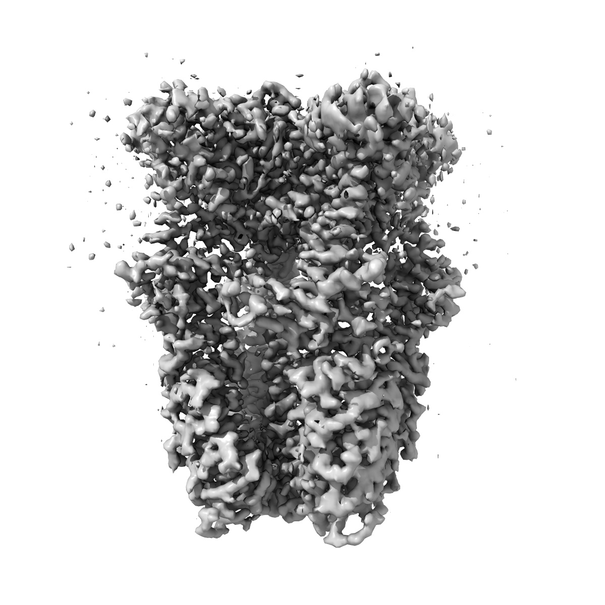
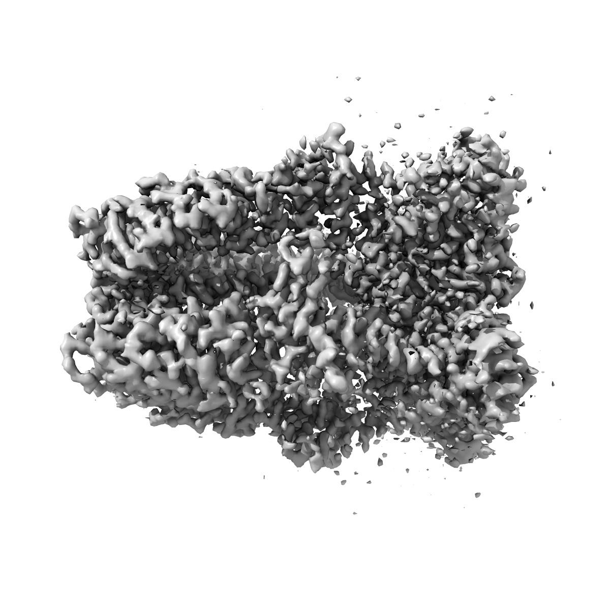
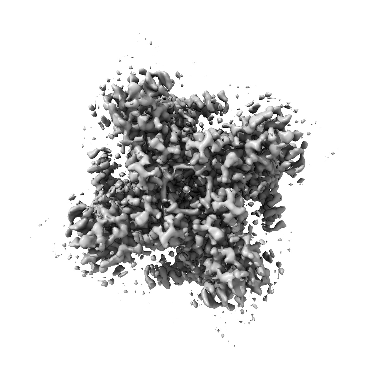
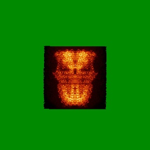
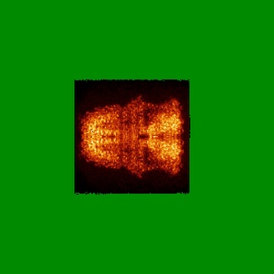
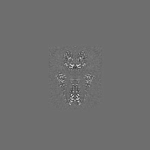
EMD-22009
Structure of human TRPA1 in complex with agonist GNE551
EMD-22009
Single-particle3.0 Å
 Deposition: 20/05/2020
Map released: 18/11/2020
Last modified: 06/03/2024
Concentration: 0.33
mg/mL
Buffer
Grid
Vitrification
Cryogen name: ETHANE
Chamber humidity: 100%
Chamber temperature: 277 K
Instrument: FEI VITROBOT MARK IV
Details: Triple blot. Put blot in vitroblot Apply 3.5ul to grid, wait 30sec, blot manually Apply 3.5ul to grid, wait 30sec, blot manually, apply final 3.5ul, final blot by vitrobot (3.5s).
Chamber humidity: 100%
Chamber temperature: 277 K
Instrument: FEI VITROBOT MARK IV
Details: Triple blot. Put blot in vitroblot Apply 3.5ul to grid, wait 30sec, blot manually Apply 3.5ul to grid, wait 30sec, blot manually, apply final 3.5ul, final blot by vitrobot (3.5s).
Microscope: FEI TITAN KRIOS
Illumination mode: FLOOD BEAM
Imaging mode: BRIGHT FIELD
Electron source: FIELD EMISSION GUN
Acceleration voltage: 300 kV
C2 aperture diameter: 70.0 µm
Nominal CS: 2.7 mm
Nominal magnification: 165000.0
Specimen holder model: FEI TITAN KRIOS AUTOGRID HOLDER
Cooling holder cryogen: NITROGEN
Alignment procedure: COMA FREE
Specialist optics
Energy filter
Image Recording
[1]
Detector model:
GATAN K2 QUANTUM (4k x 4k)
Detector mode: COUNTING
Frames per image: 1-40
Number of grids: 1
Number of real images: 11063
Average exposure time: 1.6 s
Average electron dose per image: 41.8 e/Å2
Detector mode: COUNTING
Frames per image: 1-40
Number of grids: 1
Number of real images: 11063
Average exposure time: 1.6 s
Average electron dose per image: 41.8 e/Å2
Final
reconstruction
Resolution: 3.0
Å
(
BY AUTHOR)
Resolution method: FSC 0.143 CUT-OFF
Number of images used: 58837
Algorithm: FOURIER SPACE
Resolution method: FSC 0.143 CUT-OFF
Number of images used: 58837
Algorithm: FOURIER SPACE
⌯ Applied Symmetry
Point group:
C4
Software
[1]
| Name | Version | Details |
|---|---|---|
| CisTEM | - | - |
Startup model
[1]
Type:
NONE
⦨ Initial angle
assignment
Particle selection
[1]
| Selected | Ref. model | Method | Software | Details |
|---|---|---|---|---|
| 505112 | - | - | - | - |
Format: CCP4
Data type: IMAGE STORED AS FLOATING POINT NUMBER (4 BYTES)
Annotation details: Density-modified map
Data type: IMAGE STORED AS FLOATING POINT NUMBER (4 BYTES)
Annotation details: Density-modified map
⬡ Geometry
| X | Y | Z | |
|---|---|---|---|
| Dimensions | 324 | 324 | 324 |
| Origin | 0 | 0 | 0 |
| Spacing | 324 | 324 | 324 |
| Voxel size | 1.0 Å | 1.0 Å | 1.0 Å |
Contour list
| Primary | Level | Source |
|---|---|---|
| True | 0.2 | AUTHOR |