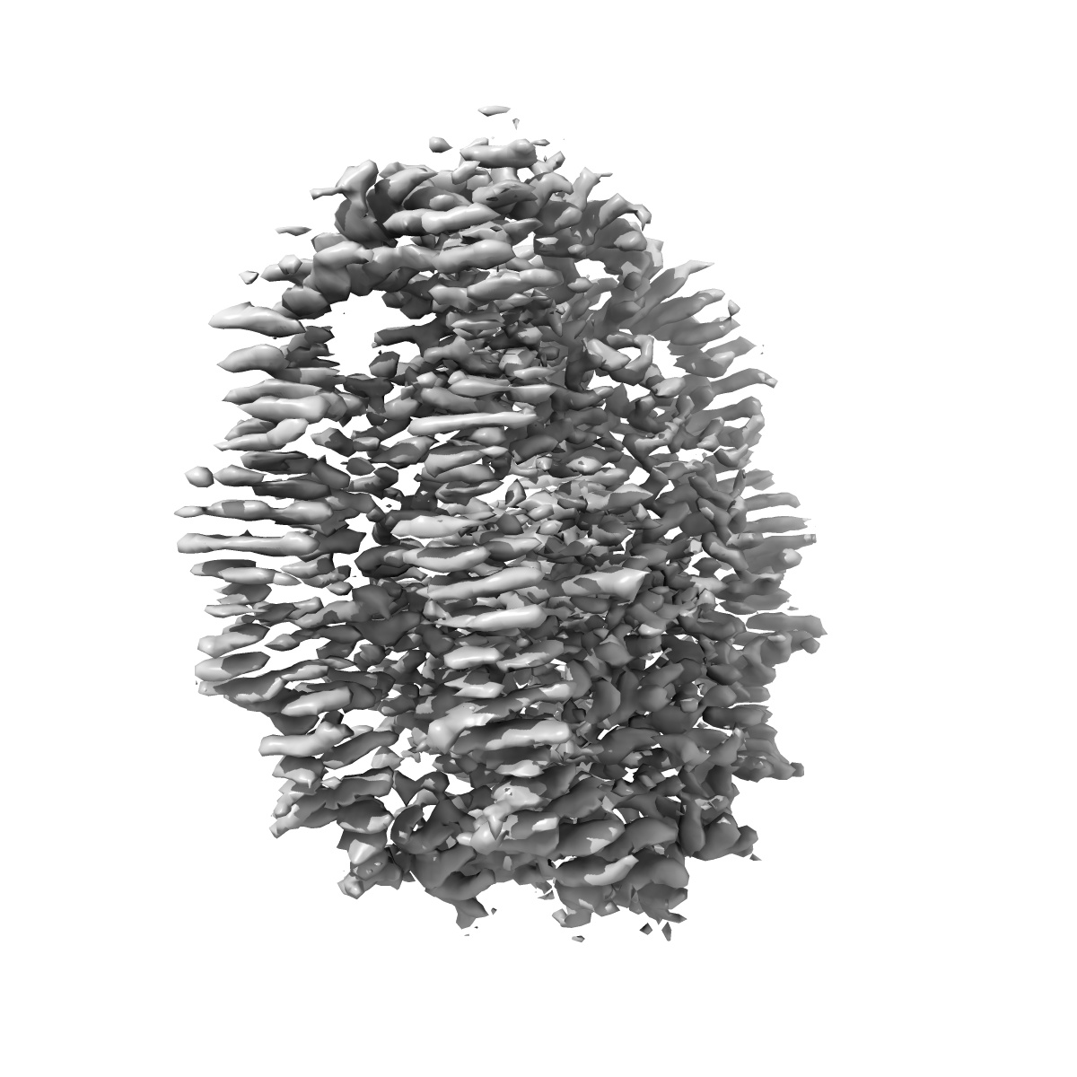
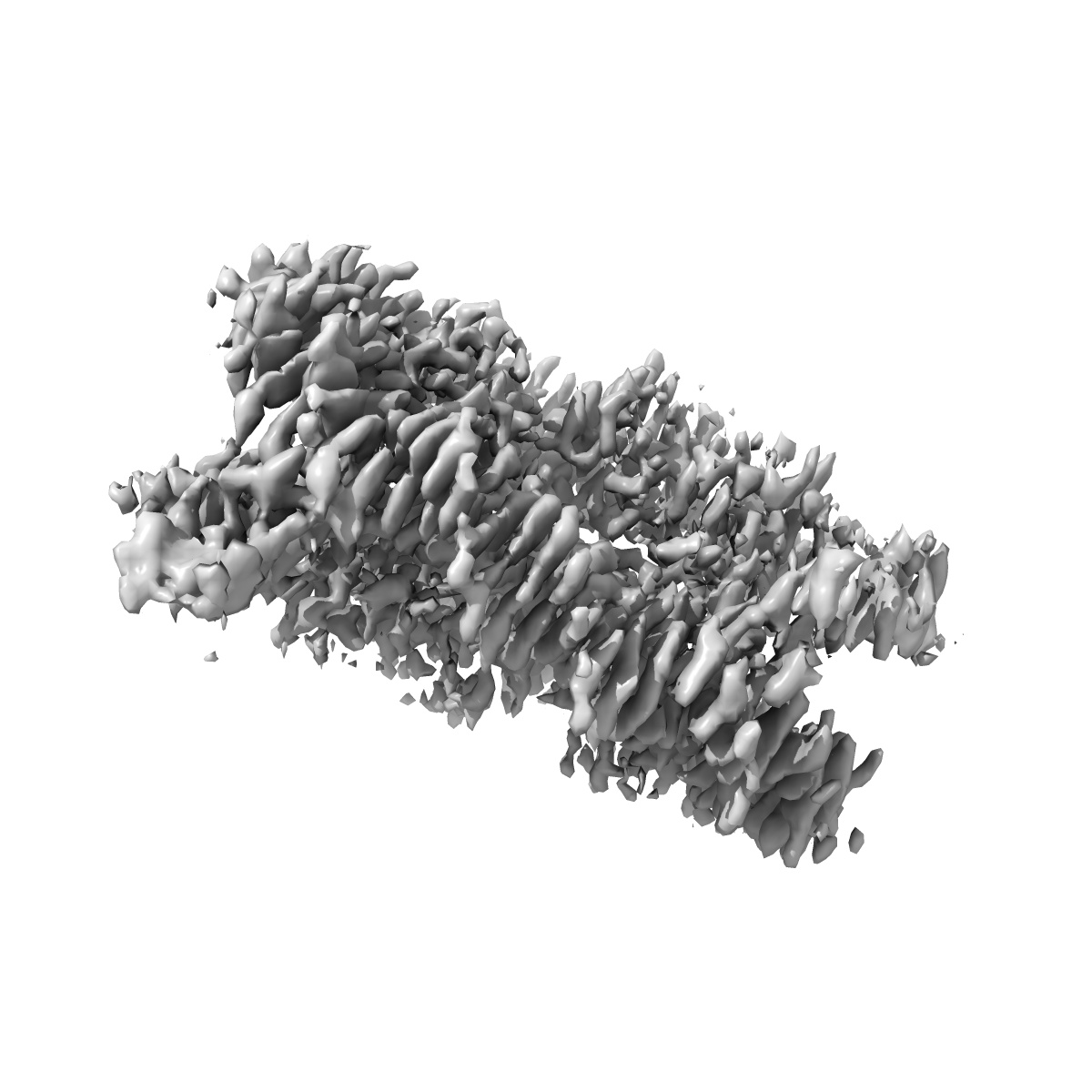
EMD-21971
Bridging of double-strand DNA break activates PARP2/HPF1 to modify chromatin
EMD-21971
Single-particle2.8 Å
 Deposition: 13/05/2020
Map released: 16/09/2020
Last modified: 06/03/2024
Microscope: FEI TITAN KRIOS
Illumination mode: FLOOD BEAM
Imaging mode: BRIGHT FIELD
Electron source: FIELD EMISSION GUN
Acceleration voltage: 300 kV
Illumination mode: FLOOD BEAM
Imaging mode: BRIGHT FIELD
Electron source: FIELD EMISSION GUN
Acceleration voltage: 300 kV
Image Recording
[1]
Detector model:
GATAN K3 BIOQUANTUM (6k x 4k)
Detector mode: COUNTING
Average electron dose per image: 80.0 e/Å2
Detector mode: COUNTING
Average electron dose per image: 80.0 e/Å2
Final
reconstruction
Resolution: 2.8
Å
(
BY AUTHOR)
Resolution method: FSC 0.143 CUT-OFF
Number of images used: 934000
Resolution method: FSC 0.143 CUT-OFF
Number of images used: 934000
⌯ Applied Symmetry
Point group:
C2
Startup model
[1]
Type:
EMDB MAP
⦨ Initial angle
assignment
Type:
OTHER
⦩ Final angle assignment
Type:
MAXIMUM LIKELIHOOD
Format: CCP4
Data type: IMAGE STORED AS FLOATING POINT NUMBER (4 BYTES)
Annotation details: Nucleosome 1
Data type: IMAGE STORED AS FLOATING POINT NUMBER (4 BYTES)
Annotation details: Nucleosome 1
⬡ Geometry
| X | Y | Z | |
|---|---|---|---|
| Dimensions | 480 | 480 | 480 |
| Origin | 0 | 0 | 0 |
| Spacing | 480 | 480 | 480 |
| Voxel size | 1.06 Å | 1.06 Å | 1.06 Å |
Contour list
| Primary | Level | Source |
|---|---|---|
| True | 0.04 | AUTHOR |