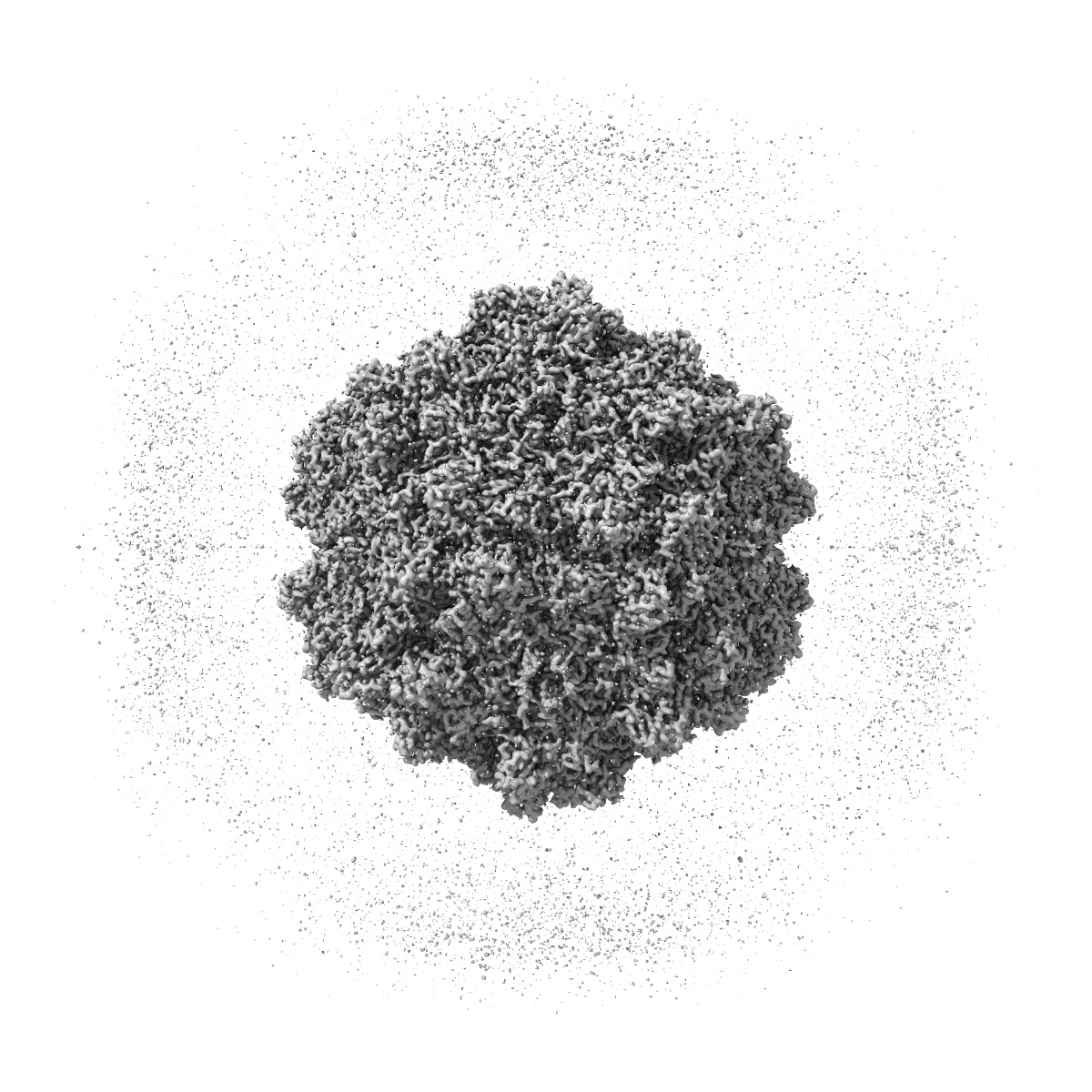
EMD-21656
BatAAV-10HB - genome-containing particles
EMD-21656
Single-particle3.03 Å
 Deposition: 04/04/2020
Map released: 24/06/2020
Last modified: 06/03/2024
Buffer
pH: 7.4
Vitrification
Cryogen name: ETHANE
Microscope: FEI TITAN KRIOS
Illumination mode: FLOOD BEAM
Imaging mode: BRIGHT FIELD
Electron source: FIELD EMISSION GUN
Acceleration voltage: 300 kV
Illumination mode: FLOOD BEAM
Imaging mode: BRIGHT FIELD
Electron source: FIELD EMISSION GUN
Acceleration voltage: 300 kV
Image Recording
[1]
Final
reconstruction
Resolution: 3.03
Å
(
BY AUTHOR)
Resolution method: FSC 0.143 CUT-OFF
Number of images used: 4661
Resolution method: FSC 0.143 CUT-OFF
Number of images used: 4661
Software
[1]
| Name | Version | Details |
|---|---|---|
| CisTEM | - | - |
Startup model
[1]
Type:
OTHER
⦨ Initial angle
assignment
Type:
RANDOM ASSIGNMENT
⦩ Final angle assignment
Type:
RANDOM ASSIGNMENT
Format: CCP4
Data type: IMAGE STORED AS FLOATING POINT NUMBER (4 BYTES)
Data type: IMAGE STORED AS FLOATING POINT NUMBER (4 BYTES)
⬡ Geometry
| X | Y | Z | |
|---|---|---|---|
| Dimensions | 400 | 400 | 400 |
| Origin | -200 | -200 | -200 |
| Spacing | 400 | 400 | 400 |
| Voxel size | 1.064 Å | 1.064 Å | 1.064 Å |
Contour list
| Primary | Level | Source |
|---|---|---|
| True | 2.0 | AUTHOR |