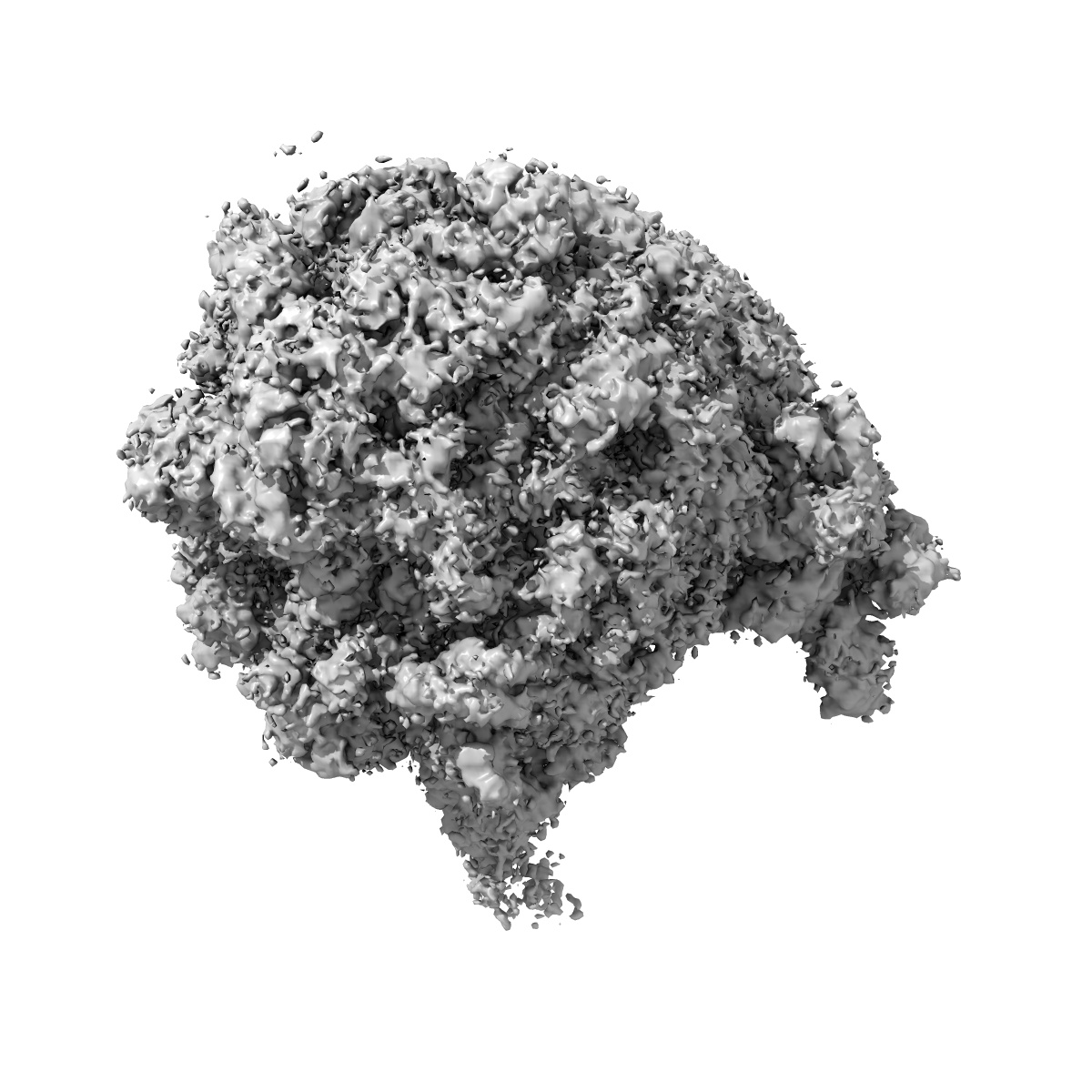
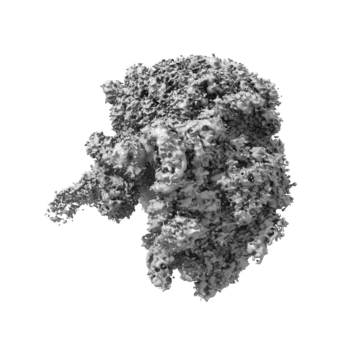
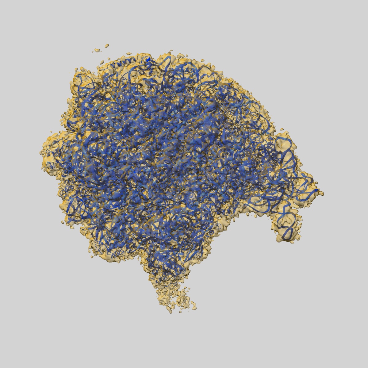
EMD-21632
Cryo-EM of elongating ribosome with EF-Tu*GTP elucidates tRNA proofreading (Cognate Structure V-A)
EMD-21632
Single-particle3.2 Å
 Deposition: 31/03/2020
Map released: 01/07/2020
Last modified: 06/03/2024
Microscope: FEI TITAN KRIOS
Illumination mode: FLOOD BEAM
Imaging mode: BRIGHT FIELD
Electron source: FIELD EMISSION GUN
Acceleration voltage: 300 kV
Illumination mode: FLOOD BEAM
Imaging mode: BRIGHT FIELD
Electron source: FIELD EMISSION GUN
Acceleration voltage: 300 kV
Image Recording
[1]
Detector model:
GATAN K2 SUMMIT (4k x 4k)
Detector mode: SUPER-RESOLUTION
Frames per image: 1-35
Number of grids: 1
Number of real images: 3218
Average exposure time: 1.0 s
Average electron dose per image: 35.0 e/Å2
Detector mode: SUPER-RESOLUTION
Frames per image: 1-35
Number of grids: 1
Number of real images: 3218
Average exposure time: 1.0 s
Average electron dose per image: 35.0 e/Å2
Final
reconstruction
Resolution: 3.2
Å
(
BY AUTHOR)
Resolution method: FSC 0.143 CUT-OFF
Number of images used: 32253
Resolution method: FSC 0.143 CUT-OFF
Number of images used: 32253
⌯ Applied Symmetry
Point group:
C1
Software
[1]
| Name | Version | Details |
|---|---|---|
| FREALIGN | - | - |
⦨ Initial angle
assignment
⦩ Final angle assignment
Particle selection
[1]
| Selected | Ref. model | Method | Software | Details |
|---|---|---|---|---|
| 678268 | - | - | - | - |
Final 3D classification
Software
[1]
| Name | Version | Details |
|---|---|---|
| FREALIGN | - | - |
Format: CCP4
Data type: IMAGE STORED AS FLOATING POINT NUMBER (4 BYTES)
Annotation details: Map V-A resolution filtered with blocfilt with B-factor of -50 A2 applied
Data type: IMAGE STORED AS FLOATING POINT NUMBER (4 BYTES)
Annotation details: Map V-A resolution filtered with blocfilt with B-factor of -50 A2 applied
⬡ Geometry
| X | Y | Z | |
|---|---|---|---|
| Dimensions | 288 | 288 | 288 |
| Origin | 0 | 0 | 0 |
| Spacing | 288 | 288 | 288 |
| Voxel size | 1.333 Å | 1.333 Å | 1.333 Å |
Contour list
| Primary | Level | Source |
|---|---|---|
| True | 1.0 | AUTHOR |