EMD-21035
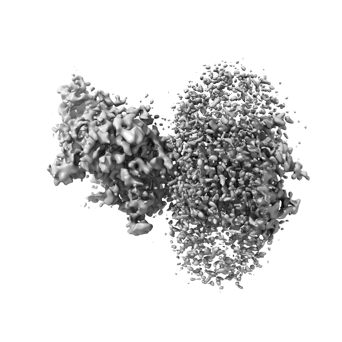
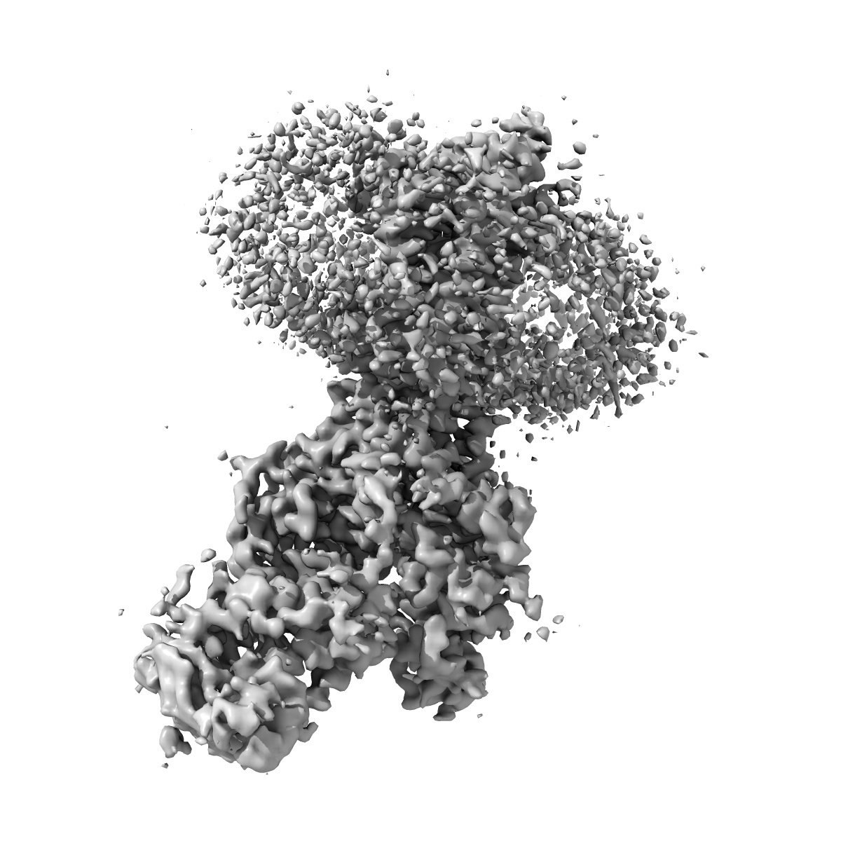
Structure of NPC1-like intracellular cholesterol transporter 1 (NPC1L1)
EMD-21035
Single-particle3.7 Å
 Deposition: 25/11/2019
Map released: 01/07/2020
Last modified: 29/07/2020
Concentration: 5
mg/mL
Buffer
pH: 7.5
Grid
Vitrification
Microscope: FEI TITAN KRIOS
Illumination mode: FLOOD BEAM
Imaging mode: BRIGHT FIELD
Electron source: FIELD EMISSION GUN
Acceleration voltage: 300 kV
C2 aperture diameter: 70.0 µm
Nominal CS: 2.7 mm
Specimen holder model: FEI TITAN KRIOS AUTOGRID HOLDER
Cooling holder cryogen: NITROGEN
Illumination mode: FLOOD BEAM
Imaging mode: BRIGHT FIELD
Electron source: FIELD EMISSION GUN
Acceleration voltage: 300 kV
C2 aperture diameter: 70.0 µm
Nominal CS: 2.7 mm
Specimen holder model: FEI TITAN KRIOS AUTOGRID HOLDER
Cooling holder cryogen: NITROGEN
Image Recording
[1]
Final
reconstruction
Resolution: 3.7
Å
(
BY AUTHOR)
Resolution method: FSC 0.143 CUT-OFF
Number of images used: 144908
Resolution method: FSC 0.143 CUT-OFF
Number of images used: 144908
⌯ Applied Symmetry
Point group:
C1
Software
[1]
| Name | Version | Details |
|---|---|---|
| RELION | 3 | - |
⦨ Initial angle
assignment
⦩ Final angle assignment
CTF correction
Software
[1]
| Name | Version | Details |
|---|---|---|
| Gctf | 1.06 | - |
Format: CCP4
Data type: IMAGE STORED AS FLOATING POINT NUMBER (4 BYTES)
Annotation details: Structure of full-length NPC1L1
Data type: IMAGE STORED AS FLOATING POINT NUMBER (4 BYTES)
Annotation details: Structure of full-length NPC1L1
⬡ Geometry
| X | Y | Z | |
|---|---|---|---|
| Dimensions | 320 | 320 | 320 |
| Origin | 0 | 0 | 0 |
| Spacing | 320 | 320 | 320 |
| Voxel size | 0.862 Å | 0.862 Å | 0.862 Å |
Contour list
| Primary | Level | Source |
|---|---|---|
| True | 0.011 | AUTHOR |