EMD-20533
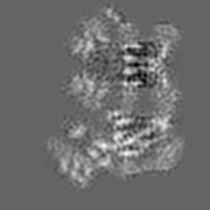
Cryo-EM structure of the pancreatic beta-cell SUR1 Apo state
EMD-20533
Single-particle4.55 Å
 Deposition: 31/07/2019
Map released: 14/08/2019
Last modified: 07/10/2020
Microscope: FEI TITAN KRIOS
Illumination mode: FLOOD BEAM
Imaging mode: BRIGHT FIELD
Electron source: FIELD EMISSION GUN
Acceleration voltage: 300 kV
Illumination mode: FLOOD BEAM
Imaging mode: BRIGHT FIELD
Electron source: FIELD EMISSION GUN
Acceleration voltage: 300 kV
Image Recording
[1]
Final
reconstruction
Resolution: 4.55
Å
(
BY AUTHOR)
Resolution method: FSC 0.143 CUT-OFF
Number of images used: 90058
Resolution method: FSC 0.143 CUT-OFF
Number of images used: 90058
⌯ Applied Symmetry
Point group:
C1
Software
[1]
| Name | Version | Details |
|---|---|---|
| RELION | - | - |
⦨ Initial angle
assignment
Type:
COMMON LINE
⦩ Final angle assignment
Type:
ANGULAR RECONSTITUTION
Format: CCP4
Data type: IMAGE STORED AS FLOATING POINT NUMBER (4 BYTES)
Annotation details: SUR1-Apo state
Data type: IMAGE STORED AS FLOATING POINT NUMBER (4 BYTES)
Annotation details: SUR1-Apo state
⬡ Geometry
| X | Y | Z | |
|---|---|---|---|
| Dimensions | 110 | 110 | 110 |
| Origin | 20 | 41 | 30 |
| Spacing | 110 | 110 | 110 |
| Voxel size | 1.826 Å | 1.826 Å | 1.826 Å |
Contour list
| Primary | Level | Source |
|---|---|---|
| True | 0.0328 | AUTHOR |