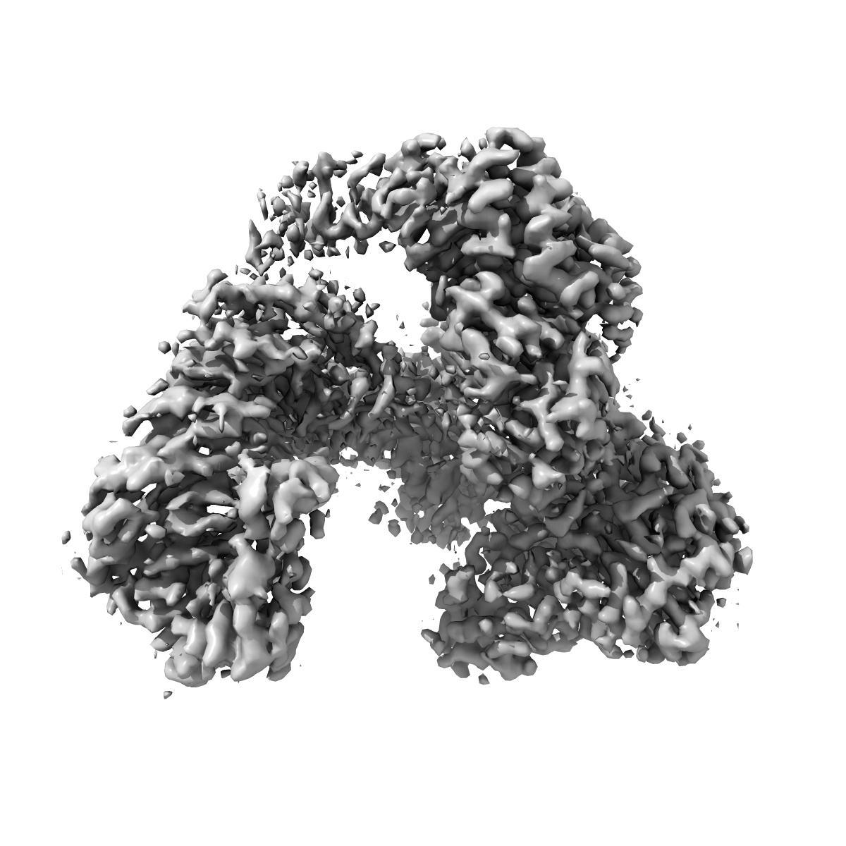
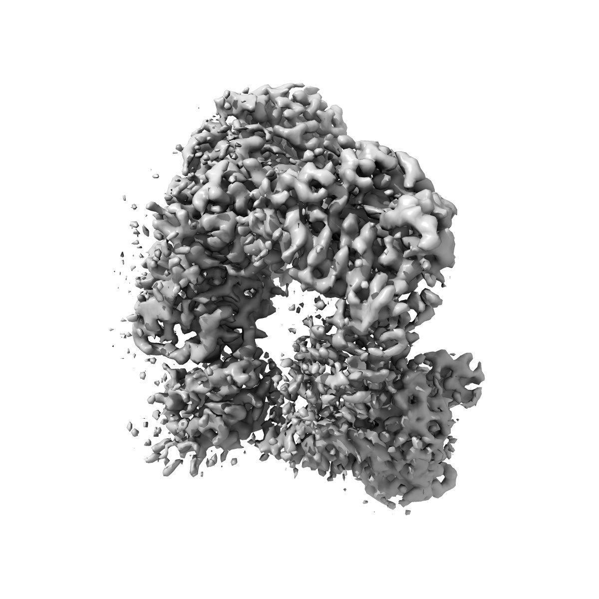
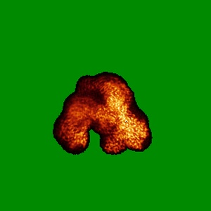
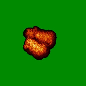
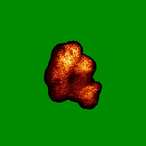
EMD-20049
CryoEM focus classification map of the hyperactive ClpB mutant K476C, bound to casein, pre-state
EMD-20049
Single-particle3.3 Å
 Deposition: 01/04/2019
Map released: 12/06/2019
Last modified: 20/03/2024
Microscope: FEI TITAN KRIOS
Illumination mode: FLOOD BEAM
Imaging mode: BRIGHT FIELD
Electron source: FIELD EMISSION GUN
Acceleration voltage: 300 kV
C2 aperture diameter: 2.6 µm
Illumination mode: FLOOD BEAM
Imaging mode: BRIGHT FIELD
Electron source: FIELD EMISSION GUN
Acceleration voltage: 300 kV
C2 aperture diameter: 2.6 µm
Image Recording
[1]
Final
reconstruction
Resolution: 3.3
Å
(
BY AUTHOR)
Resolution method: FSC 0.143 CUT-OFF
Number of images used: 221083
Resolution method: FSC 0.143 CUT-OFF
Number of images used: 221083
⌯ Applied Symmetry
Point group:
C1
Startup model
[1]
Type:
INSILICO MODEL
⦨ Initial angle
assignment
Type:
MAXIMUM LIKELIHOOD
⦩ Final angle assignment
Type:
MAXIMUM LIKELIHOOD
Format: CCP4
Data type: IMAGE STORED AS FLOATING POINT NUMBER (4 BYTES)
Annotation details: EM focus classification map of the hyperactive ClpB mutant K476C, bound to casein, pre-state
Data type: IMAGE STORED AS FLOATING POINT NUMBER (4 BYTES)
Annotation details: EM focus classification map of the hyperactive ClpB mutant K476C, bound to casein, pre-state
⬡ Geometry
| X | Y | Z | |
|---|---|---|---|
| Dimensions | 256 | 256 | 256 |
| Origin | 0 | 0 | 0 |
| Spacing | 256 | 256 | 256 |
| Voxel size | 1.032 Å | 1.032 Å | 1.032 Å |
Contour list
| Primary | Level | Source |
|---|---|---|
| True | 0.0464 | AUTHOR |