EMD-20002
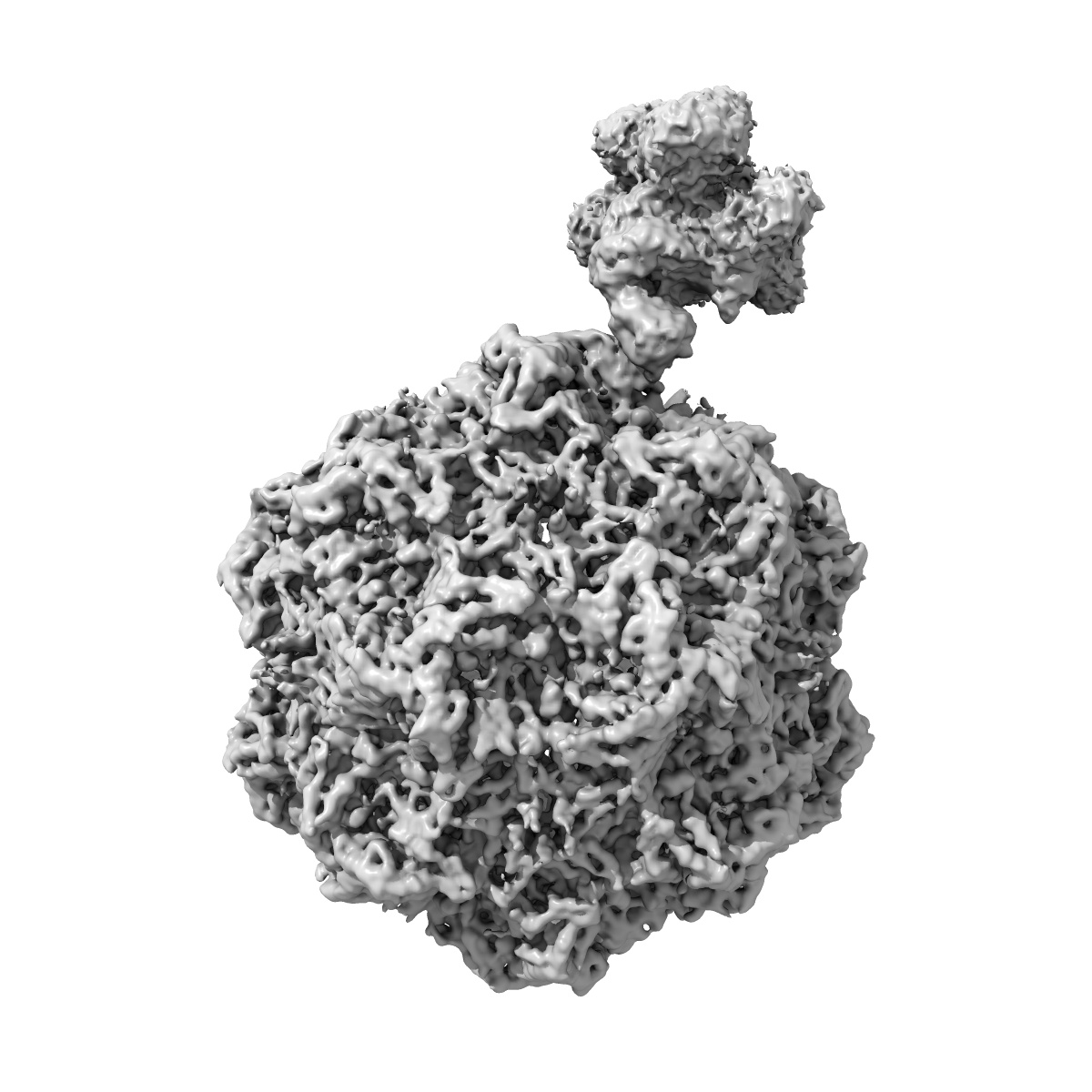
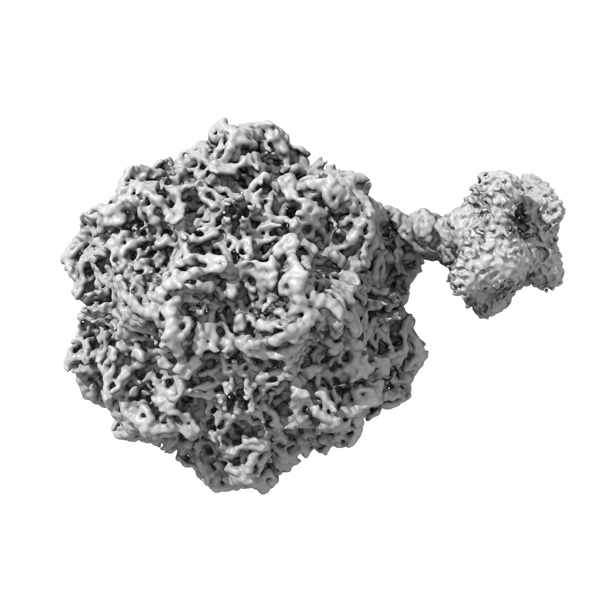
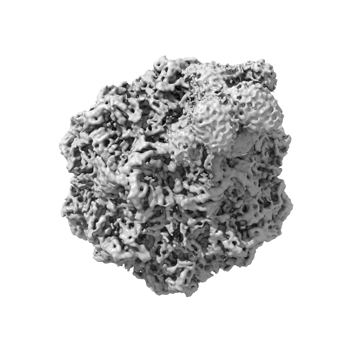
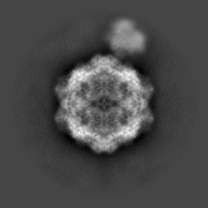
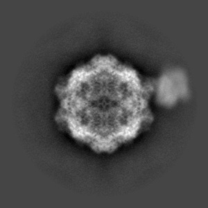
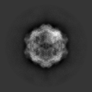
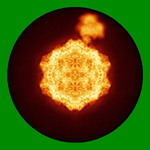
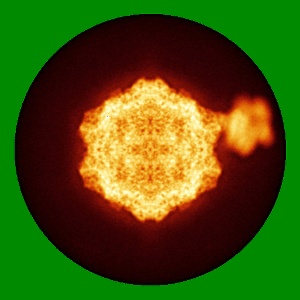
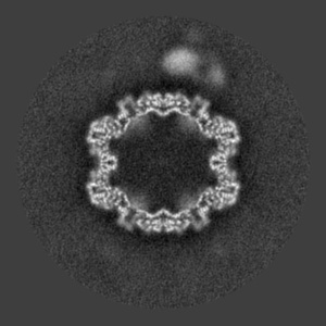
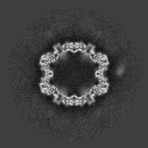
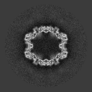
Structure of canine parvovirus in complex with transferrin receptor type-1
EMD-20002
Single-particle6.2 Å
 Deposition: 18/03/2019
Map released: 18/09/2019
Last modified: 23/10/2019
Microscope: FEI TITAN KRIOS
Illumination mode: SPOT SCAN
Imaging mode: BRIGHT FIELD
Electron source: FIELD EMISSION GUN
Acceleration voltage: 300 kV
Illumination mode: SPOT SCAN
Imaging mode: BRIGHT FIELD
Electron source: FIELD EMISSION GUN
Acceleration voltage: 300 kV
Image Recording
[1]
Final
reconstruction
Resolution: 6.2
Å
(
BY AUTHOR)
Resolution method: FSC 0.143 CUT-OFF
Number of images used: 62005
Resolution method: FSC 0.143 CUT-OFF
Number of images used: 62005
⌯ Applied Symmetry
Point group:
C1
⦨ Initial angle
assignment
Type:
MAXIMUM LIKELIHOOD
⦩ Final angle assignment
Type:
MAXIMUM LIKELIHOOD
Format: CCP4
Data type: IMAGE STORED AS FLOATING POINT NUMBER (4 BYTES)
Annotation details: Asymmetric map of the canine parvovirus in complex with TfR-Tf (single orientation)
Data type: IMAGE STORED AS FLOATING POINT NUMBER (4 BYTES)
Annotation details: Asymmetric map of the canine parvovirus in complex with TfR-Tf (single orientation)
⬡ Geometry
| X | Y | Z | |
|---|---|---|---|
| Dimensions | 512 | 512 | 512 |
| Origin | -256 | -256 | -256 |
| Spacing | 512 | 512 | 512 |
| Voxel size | 1.11 Å | 1.11 Å | 1.11 Å |
Contour list
| Primary | Level | Source |
|---|---|---|
| True | 0.017 | AUTHOR |