EMD-1502
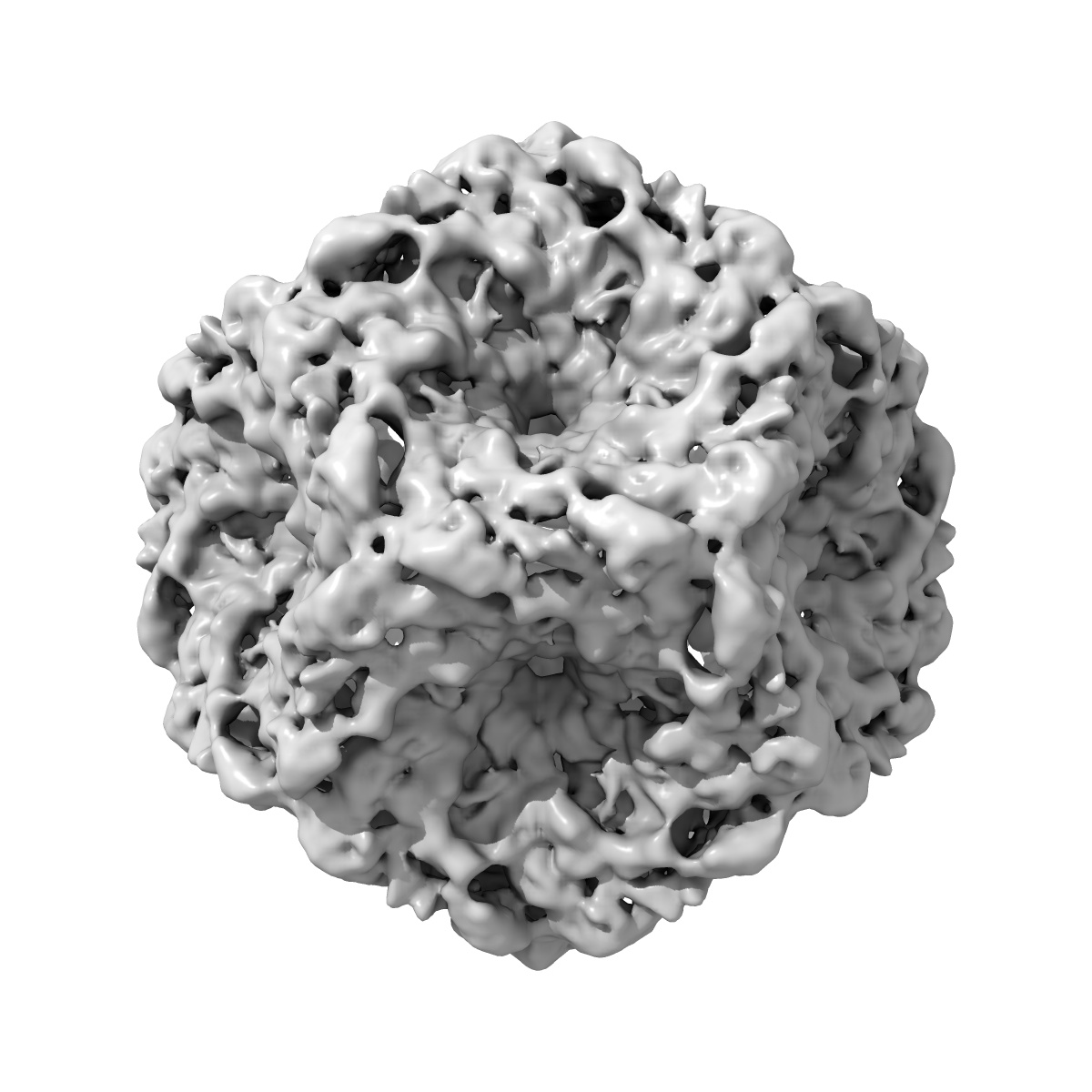
Initial location of the RNA-dependent RNA polymerase in the bacteriophage phi6 procapsid determined by cryo-electron microscopy
EMD-1502
Single-particle11.0 Å
 Deposition: 06/04/2008
Map released: 31/03/2009
Last modified: 07/11/2012
Concentration: 0.5
mg/mL
Buffer
pH: 7.5
Details: 10mM sodium phosphate, 1mM MgCl2, 150 mM NaCl
Details: 10mM sodium phosphate, 1mM MgCl2, 150 mM NaCl
Staining
Type:
NEGATIVE
Details: None
Details: None
Vitrification
Cryogen name: ETHANE
Instrument: REICHERT-JUNG PLUNGER
Details: Vitrification instrument: Reichert-Jung cryo-fixation unit
Instrument: REICHERT-JUNG PLUNGER
Details: Vitrification instrument: Reichert-Jung cryo-fixation unit
Microscope: FEI/PHILIPS CM200FEG
Illumination mode: FLOOD BEAM
Imaging mode: BRIGHT FIELD
Electron source: FIELD EMISSION GUN
Acceleration voltage: 120 kV
Nominal CS: 2 mm
Nominal defocus: 1.0 µm - 2.3 µm
Nominal magnification: 50000.0
Specimen holder model: GATAN LIQUID NITROGEN
Specimen holder details: Side entry, Eucentric
Alignment procedure: LEGACY (Astigmatism: Corrected at 300000 times magnification, Electron beam tilt params: )
Minimum tilt angle: 0
Maximum tilt angle: 0
Image Recording
[1]
Scanner:
NIKON SUPER COOLSCAN 9000
Sampling interval: 6.35 µm
Number of real images: 36
Average electron dose per image: 15 e/Å2
Bits per pixel: 16.0
Sampling interval: 6.35 µm
Number of real images: 36
Average electron dose per image: 15 e/Å2
Bits per pixel: 16.0
Final
reconstruction
Resolution: 11.0
Å
(
BY AUTHOR)
Resolution method: FSC 0.33 CUT-OFF
Number of images used: 1072
Algorithm: OTHER
Resolution method: FSC 0.33 CUT-OFF
Number of images used: 1072
Algorithm: OTHER
⌯ Applied Symmetry
Point group:
I
Software
[1]
| Name | Version | Details |
|---|---|---|
| Bsoft, PFT3DR | - | - |
CTF correction
Details:Each particle phase flipped
Format: CCP4
Data type: IMAGE STORED AS FLOATING POINT NUMBER (4 BYTES)
Annotation details: Map of a procapsid without the P2 protein, containing only proteins P1, P4 and P7
Details: ::::EMDATABANK.org::::EMD-1502::::
Data type: IMAGE STORED AS FLOATING POINT NUMBER (4 BYTES)
Annotation details: Map of a procapsid without the P2 protein, containing only proteins P1, P4 and P7
Details: ::::EMDATABANK.org::::EMD-1502::::
⬡ Geometry
| X | Y | Z | |
|---|---|---|---|
| Dimensions | 234 | 234 | 234 |
| Origin | -117 | -117 | -117 |
| Spacing | 234 | 234 | 234 |
| Voxel size | 2.54 Å | 2.54 Å | 2.54 Å |
Contour list
| Primary | Level | Source |
|---|---|---|
| True | 2.25 | - |