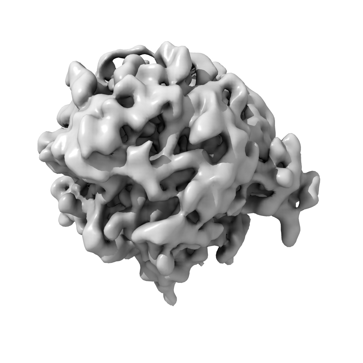
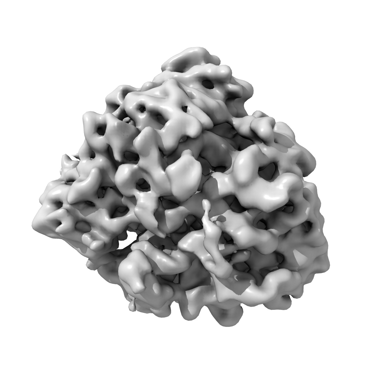
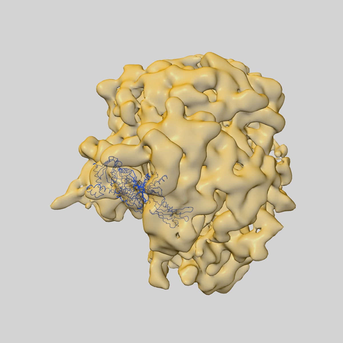
EMD-1365
Locking and unlocking of ribosomal motions.
EMD-1365
Single-particle11.75 Å
 Deposition: 21/05/2007
Map released: 24/05/2007
Last modified: 07/11/2012
Microscope: FEI TECNAI 20
Illumination mode: FLOOD BEAM
Imaging mode: BRIGHT FIELD
Electron source: FIELD EMISSION GUN
Acceleration voltage: 200 kV
Nominal magnification: 50000.0
Calibrated magnification: 49696.0
Specimen holder model: GATAN LIQUID NITROGEN
Specimen holder details: Cryo transfer
Alignment procedure: LEGACY
Minimum tilt angle: 0
Maximum tilt angle: 0
Temperature
Average: 93
K
Image Recording
[1]
Detector category:
FILM
Detector model: KODAK SO-163 FILM
Scanner: ZEISS SCAI
Sampling interval: 14 µm
Detector model: KODAK SO-163 FILM
Scanner: ZEISS SCAI
Sampling interval: 14 µm
Final
reconstruction
Resolution: 11.75
Å
(
BY AUTHOR)
Resolution method: FSC 0.5 CUT-OFF
Number of images used: 1
Algorithm: OTHER
Resolution method: FSC 0.5 CUT-OFF
Number of images used: 1
Algorithm: OTHER
⌯ Applied Symmetry
Point group:
C1
Software
[1]
| Name | Version | Details |
|---|---|---|
| SPIDER, package | - | - |
CTF correction
Details:CTF correctionn of 3D map
Format: CCP4
Data type: IMAGE STORED AS FLOATING POINT NUMBER (4 BYTES)
Annotation details: Cryo-EM map of E.coli 70S ribosome
Details: ::::EMDATABANK.org::::EMD-1365::::
Data type: IMAGE STORED AS FLOATING POINT NUMBER (4 BYTES)
Annotation details: Cryo-EM map of E.coli 70S ribosome
Details: ::::EMDATABANK.org::::EMD-1365::::
⬡ Geometry
| X | Y | Z | |
|---|---|---|---|
| Dimensions | 130 | 130 | 130 |
| Origin | -65 | -65 | -65 |
| Spacing | 130 | 130 | 130 |
| Voxel size | 2.82 Å | 2.82 Å | 2.82 Å |
Contour list
| Primary | Level | Source |
|---|---|---|
| True | 55.7 | - |