EMD-11236
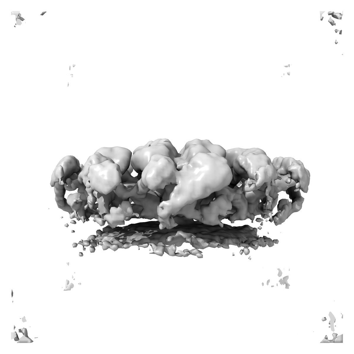
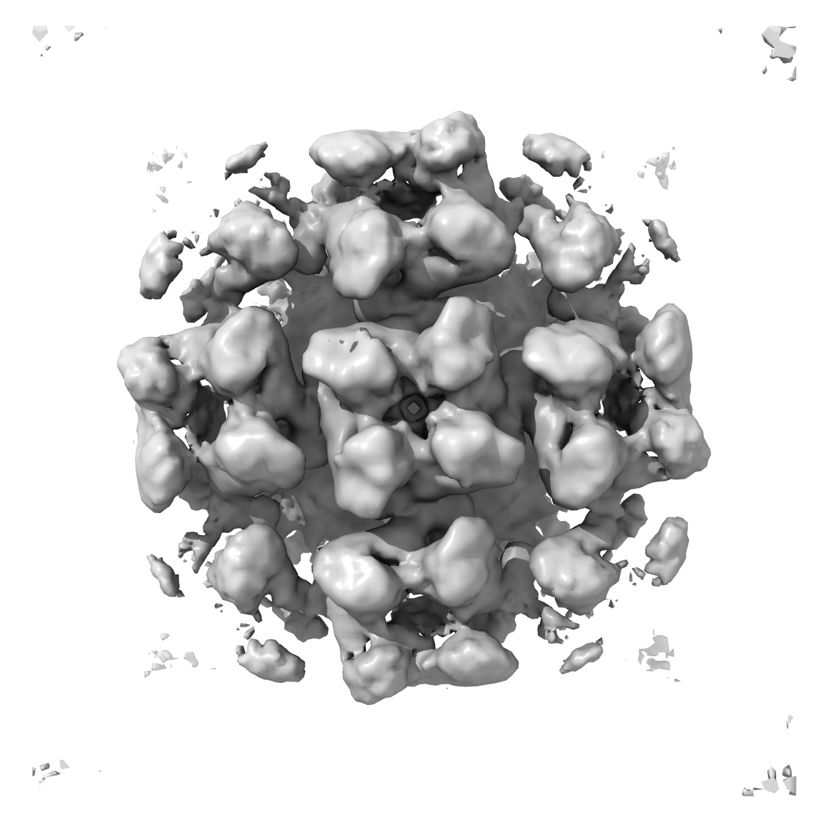
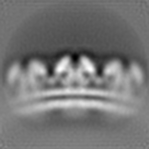
Reconstruction of Tula virus surface glycoprotein lattice
EMD-11236
Subtomogram averaging11.4 Å
 Deposition: 29/06/2020
Map released: 14/10/2020
Last modified: 28/10/2020
Buffer
pH: 7.0
Vitrification
Cryogen name: ETHANE-PROPANE
Microscope: FEI POLARA 300
Illumination mode: FLOOD BEAM
Imaging mode: BRIGHT FIELD
Electron source: FIELD EMISSION GUN
Acceleration voltage: 300 kV
Illumination mode: FLOOD BEAM
Imaging mode: BRIGHT FIELD
Electron source: FIELD EMISSION GUN
Acceleration voltage: 300 kV
Specialist optics
Energy filter
Image Recording
[1]
Detector model:
GATAN K2 SUMMIT (4k x 4k)
Detector mode: COUNTING
Average electron dose per image: 4.71 e/Å2
Detector mode: COUNTING
Average electron dose per image: 4.71 e/Å2
Final
reconstruction
Resolution: 11.4
Å
(
BY AUTHOR)
Resolution method: FSC 0.143 CUT-OFF
Resolution method: FSC 0.143 CUT-OFF
⌯ Applied Symmetry
Point group:
C4
Software
[1]
| Name | Version | Details |
|---|---|---|
| Dynamo | - | - |
⦩ Final angle assignment
Final 3D classification
Number of classes:
8
Software
[1]
| Name | Version | Details |
|---|---|---|
| RELION | - | - |
Extraction
Number of images used: 107820
Reference model: EMD-4867
Method: Template matching
Reference model: EMD-4867
Method: Template matching
Software
[1]
| Name | Version | Details |
|---|---|---|
| Dynamo | - | - |
CTF correction
Software
[1]
| Name | Version | Details |
|---|---|---|
| ETomo | - | - |
Format: CCP4
Data type: IMAGE STORED AS FLOATING POINT NUMBER (4 BYTES)
Annotation details: Reconstruction of Tula virus glycoprotein spike lattice.
Data type: IMAGE STORED AS FLOATING POINT NUMBER (4 BYTES)
Annotation details: Reconstruction of Tula virus glycoprotein spike lattice.
⬡ Geometry
| X | Y | Z | |
|---|---|---|---|
| Dimensions | 108 | 108 | 108 |
| Origin | 0 | 0 | 0 |
| Spacing | 108 | 108 | 108 |
| Voxel size | 4.0 Å | 4.0 Å | 4.0 Å |
Contour list
| Primary | Level | Source |
|---|---|---|
| True | 1.5 | AUTHOR |