EMD-11164
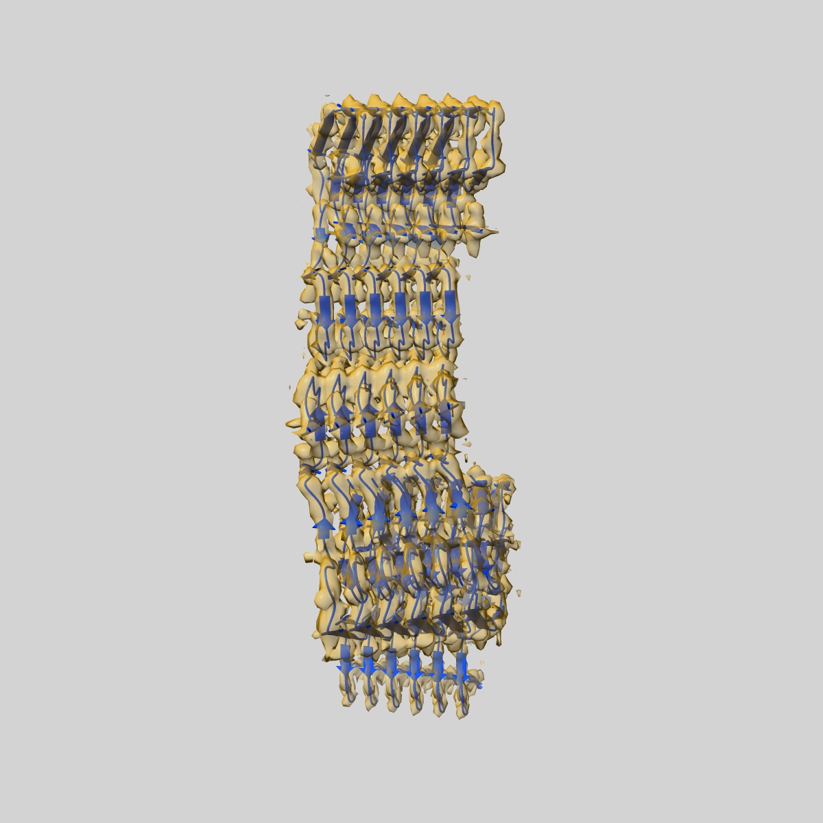
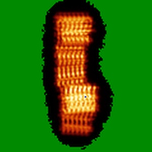
Amyloid fibril morphology II (ex vivo) from murine SAA1.1 protein.
EMD-11164
Helical reconstruction3.5 Å
 Deposition: 11/06/2020
Map released: 17/02/2021
Last modified: 24/02/2021
Buffer
pH: 7.0
Details: Water
Details: Water
Vitrification
Microscope: FEI TITAN KRIOS
Illumination mode: FLOOD BEAM
Imaging mode: BRIGHT FIELD
Electron source: FIELD EMISSION GUN
Acceleration voltage: 300 kV
Cooling holder cryogen: NITROGEN
Illumination mode: FLOOD BEAM
Imaging mode: BRIGHT FIELD
Electron source: FIELD EMISSION GUN
Acceleration voltage: 300 kV
Cooling holder cryogen: NITROGEN
Image Recording
[1]
Detector model:
GATAN K2 SUMMIT (4k x 4k)
Detector mode: COUNTING
Average electron dose per image: 20.0 e/Å2
Detector mode: COUNTING
Average electron dose per image: 20.0 e/Å2
Final
reconstruction
Resolution: 3.5
Å
(
BY AUTHOR)
Resolution method: FSC 0.143 CUT-OFF
Number of images used: 15505
Resolution method: FSC 0.143 CUT-OFF
Number of images used: 15505
⌯ Applied Symmetry
Software
[1]
| Name | Version | Details |
|---|---|---|
| RELION | 2.1 | - |
⦩ Final angle assignment
Type:
NOT APPLICABLE
CTF correction
Software
[1]
| Name | Version | Details |
|---|---|---|
| Gctf | - | - |
Segment selection
[1]
| Number selected | Segment length | Segment overlap | Total filament length | Details |
|---|---|---|---|---|
| 15530 | - | - | - | - |
Format: CCP4
Data type: IMAGE STORED AS FLOATING POINT NUMBER (4 BYTES)
Annotation details: Ex vivo mSAA amyloid fibril morphology II
Data type: IMAGE STORED AS FLOATING POINT NUMBER (4 BYTES)
Annotation details: Ex vivo mSAA amyloid fibril morphology II
⬡ Geometry
| X | Y | Z | |
|---|---|---|---|
| Dimensions | 120 | 120 | 120 |
| Origin | 0 | 0 | 0 |
| Spacing | 120 | 120 | 120 |
| Voxel size | 1.35 Å | 1.35 Å | 1.35 Å |
Contour list
| Primary | Level | Source |
|---|---|---|
| True | 13.0 | AUTHOR |