EMD-11063
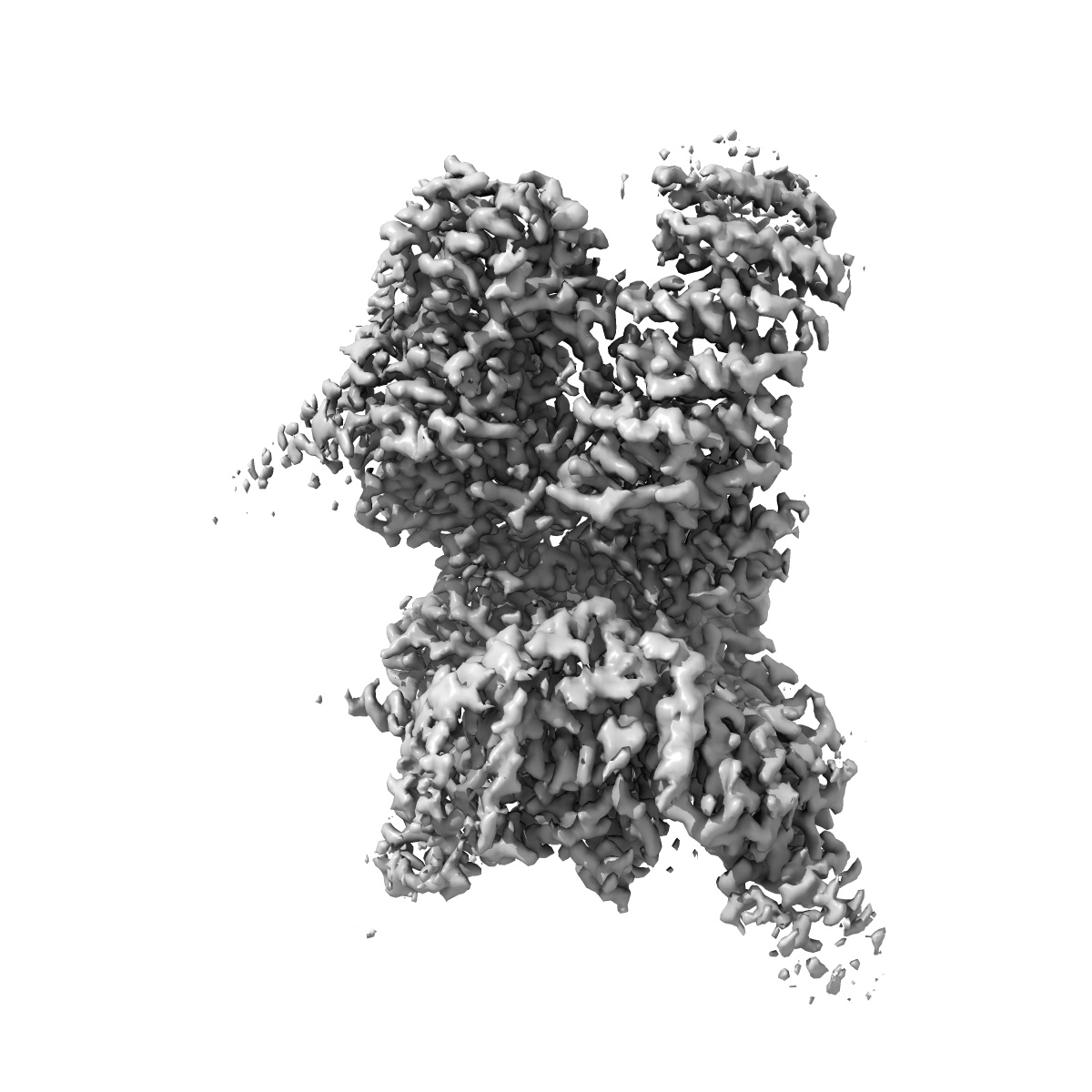
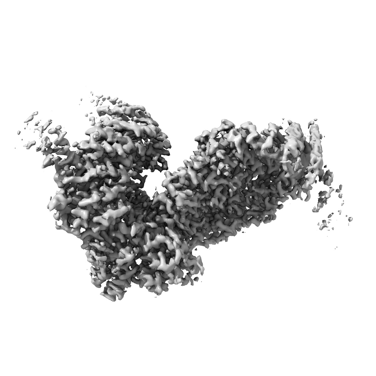
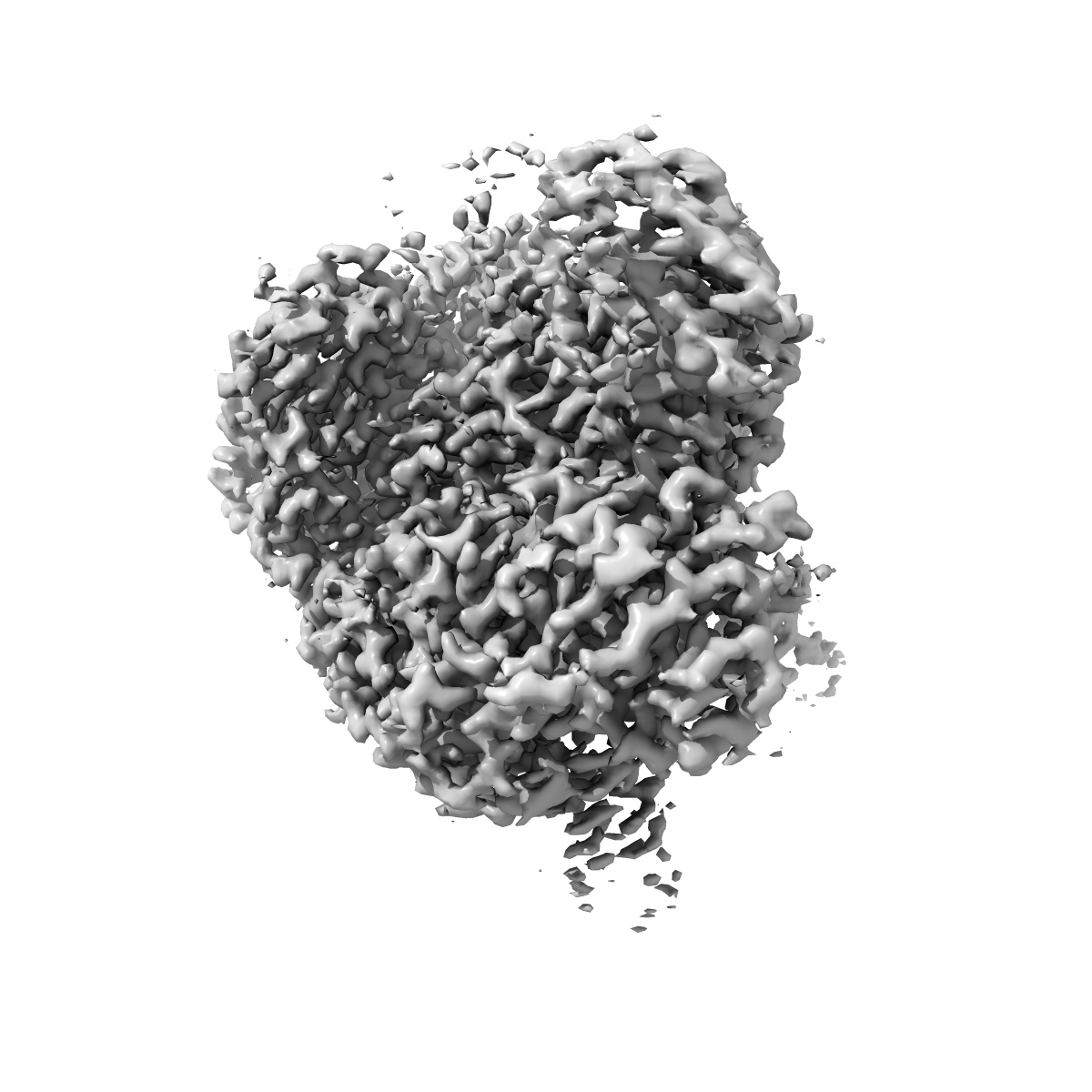
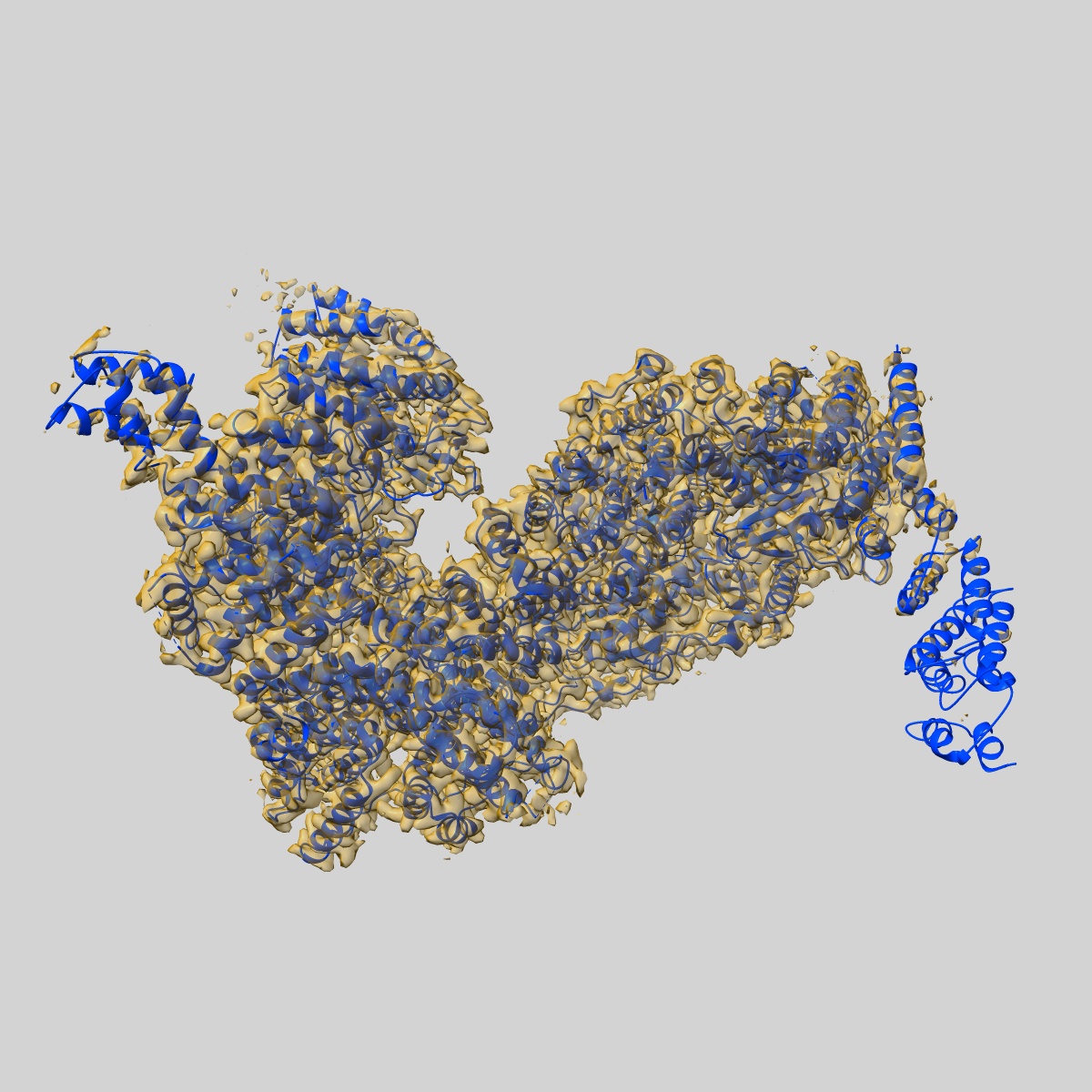
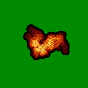
Structure of SMG1-8-9 kinase complex bound to UPF1-LSQ
EMD-11063
Single-particle2.97 Å
 Deposition: 21/05/2020
Map released: 24/06/2020
Last modified: 07/10/2020
Microscope: FEI TITAN KRIOS
Illumination mode: OTHER
Imaging mode: OTHER
Electron source: FIELD EMISSION GUN
Acceleration voltage: 300 kV
Cooling holder cryogen: NITROGEN
Illumination mode: OTHER
Imaging mode: OTHER
Electron source: FIELD EMISSION GUN
Acceleration voltage: 300 kV
Cooling holder cryogen: NITROGEN
Specialist optics
Energy filter
Image Recording
[1]
Detector model:
GATAN K3 (6k x 4k)
Number of grids: 1
Number of real images: 6293
Average exposure time: 5.5 s
Average electron dose per image: 68.75 e/Å2
Number of grids: 1
Number of real images: 6293
Average exposure time: 5.5 s
Average electron dose per image: 68.75 e/Å2
Final
reconstruction
Resolution: 2.97
Å
(
BY AUTHOR)
Resolution method: FSC 0.143 CUT-OFF
Number of images used: 481754
Resolution method: FSC 0.143 CUT-OFF
Number of images used: 481754
⌯ Applied Symmetry
Point group:
C1
Software
[1]
| Name | Version | Details |
|---|---|---|
| RELION | 3.0 | - |
⦨ Initial angle
assignment
Type:
MAXIMUM LIKELIHOOD
⦩ Final angle assignment
Type:
MAXIMUM LIKELIHOOD
Particle selection
[1]
| Selected | Ref. model | Method | Software | Details |
|---|---|---|---|---|
| 4368586 | - | - | - | - |
CTF correction
Software
[1]
| Name | Version | Details |
|---|---|---|
| CTFFIND | 4.1 | - |
Format: CCP4
Data type: IMAGE STORED AS FLOATING POINT NUMBER (4 BYTES)
Annotation details: SMG1-8-9 with bound UPF1-LSQ
Data type: IMAGE STORED AS FLOATING POINT NUMBER (4 BYTES)
Annotation details: SMG1-8-9 with bound UPF1-LSQ
⬡ Geometry
| X | Y | Z | |
|---|---|---|---|
| Dimensions | 320 | 320 | 320 |
| Origin | 0 | 0 | 0 |
| Spacing | 320 | 320 | 320 |
| Voxel size | 1.096 Å | 1.096 Å | 1.096 Å |
Contour list
| Primary | Level | Source |
|---|---|---|
| True | 0.0312 | AUTHOR |