EMD-10898
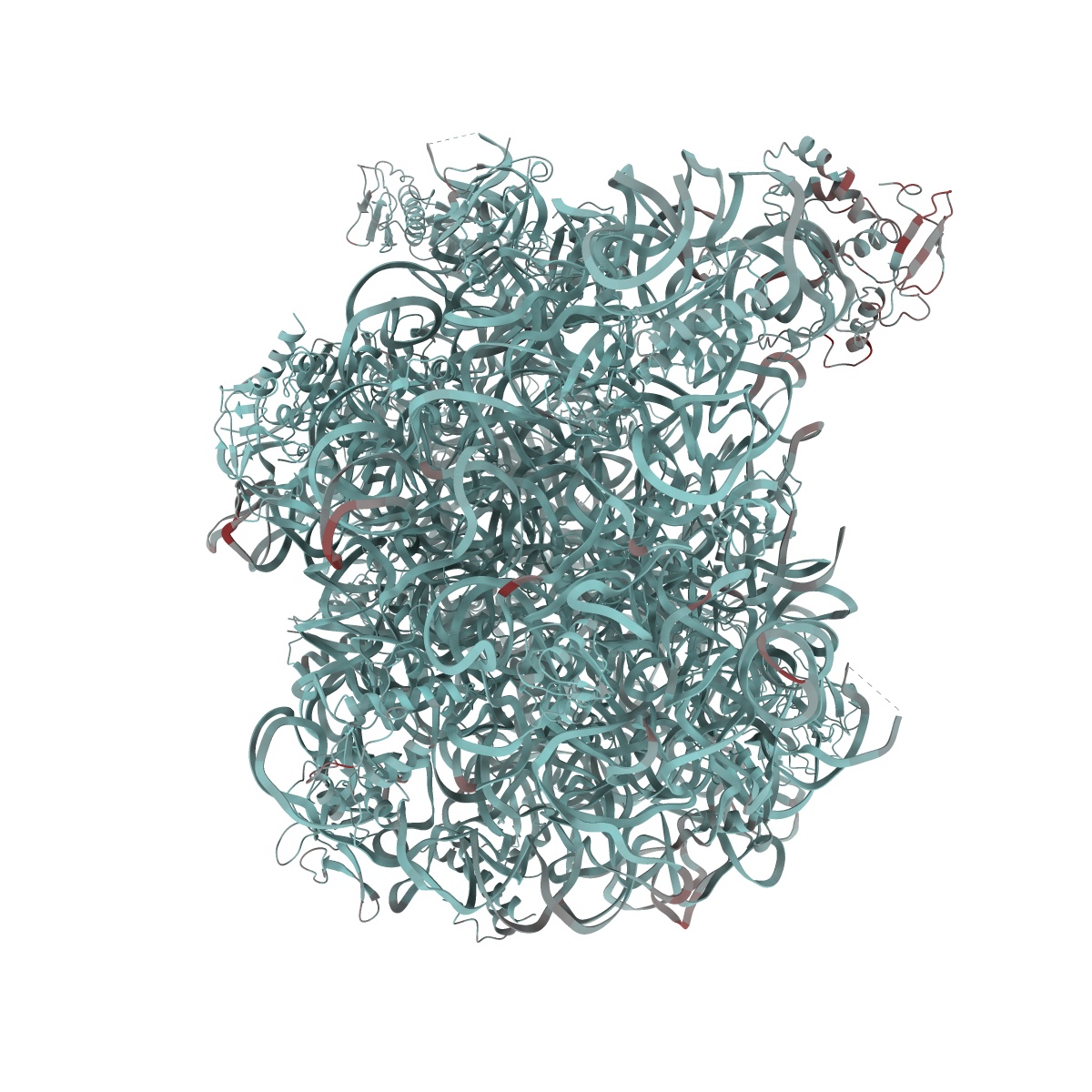
Acinetobacter baumannii ribosome-tigecycline complex - 50S subunit
EMD-10898
Single-particle2.5 Å
 Deposition: 22/04/2020
Map released: 16/09/2020
Last modified: 14/10/2020
Buffer
pH: 7.5
Vitrification
Cryogen name: ETHANE
Microscope: FEI TITAN KRIOS
Illumination mode: FLOOD BEAM
Imaging mode: BRIGHT FIELD
Electron source: FIELD EMISSION GUN
Acceleration voltage: 300 kV
Nominal defocus: 0.8 µm - 2.6 µm
Nominal magnification: 75000.0
Illumination mode: FLOOD BEAM
Imaging mode: BRIGHT FIELD
Electron source: FIELD EMISSION GUN
Acceleration voltage: 300 kV
Nominal defocus: 0.8 µm - 2.6 µm
Nominal magnification: 75000.0
Image Recording
[1]
Detector model:
FEI FALCON III (4k x 4k)
Detector mode: INTEGRATING
Average exposure time: 1.1 s
Average electron dose per image: 62.0 e/Å2
Detector mode: INTEGRATING
Average exposure time: 1.1 s
Average electron dose per image: 62.0 e/Å2
Final
reconstruction
Resolution: 2.5
Å
(
BY AUTHOR)
Resolution method: FSC 0.143 CUT-OFF
Number of images used: 231159
Resolution method: FSC 0.143 CUT-OFF
Number of images used: 231159
⌯ Applied Symmetry
Point group:
C1
Software
[1]
| Name | Version | Details |
|---|---|---|
| RELION | 3.0 | - |
Startup model
[1]
⦨ Initial angle
assignment
⦩ Final angle assignment
CTF correction
Software
[1]
| Name | Version | Details |
|---|---|---|
| Gctf | 1.18 | - |
Format: CCP4
Data type: IMAGE STORED AS FLOATING POINT NUMBER (4 BYTES)
Annotation details: Acinetobacter baumannii ribosome-tigecycline complex - 50S subunit, post-processed map
Data type: IMAGE STORED AS FLOATING POINT NUMBER (4 BYTES)
Annotation details: Acinetobacter baumannii ribosome-tigecycline complex - 50S subunit, post-processed map
⬡ Geometry
| X | Y | Z | |
|---|---|---|---|
| Dimensions | 400 | 400 | 400 |
| Origin | 0 | 0 | 0 |
| Spacing | 400 | 400 | 400 |
| Voxel size | 1.065 Å | 1.065 Å | 1.065 Å |
Contour list
| Primary | Level | Source |
|---|---|---|
| True | 0.05 | AUTHOR |