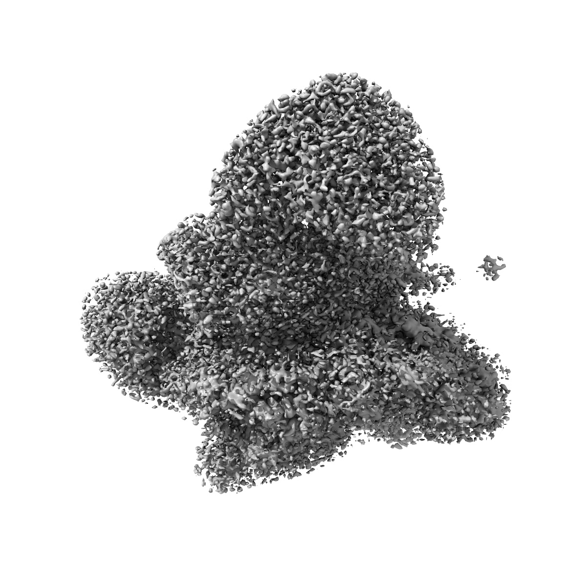
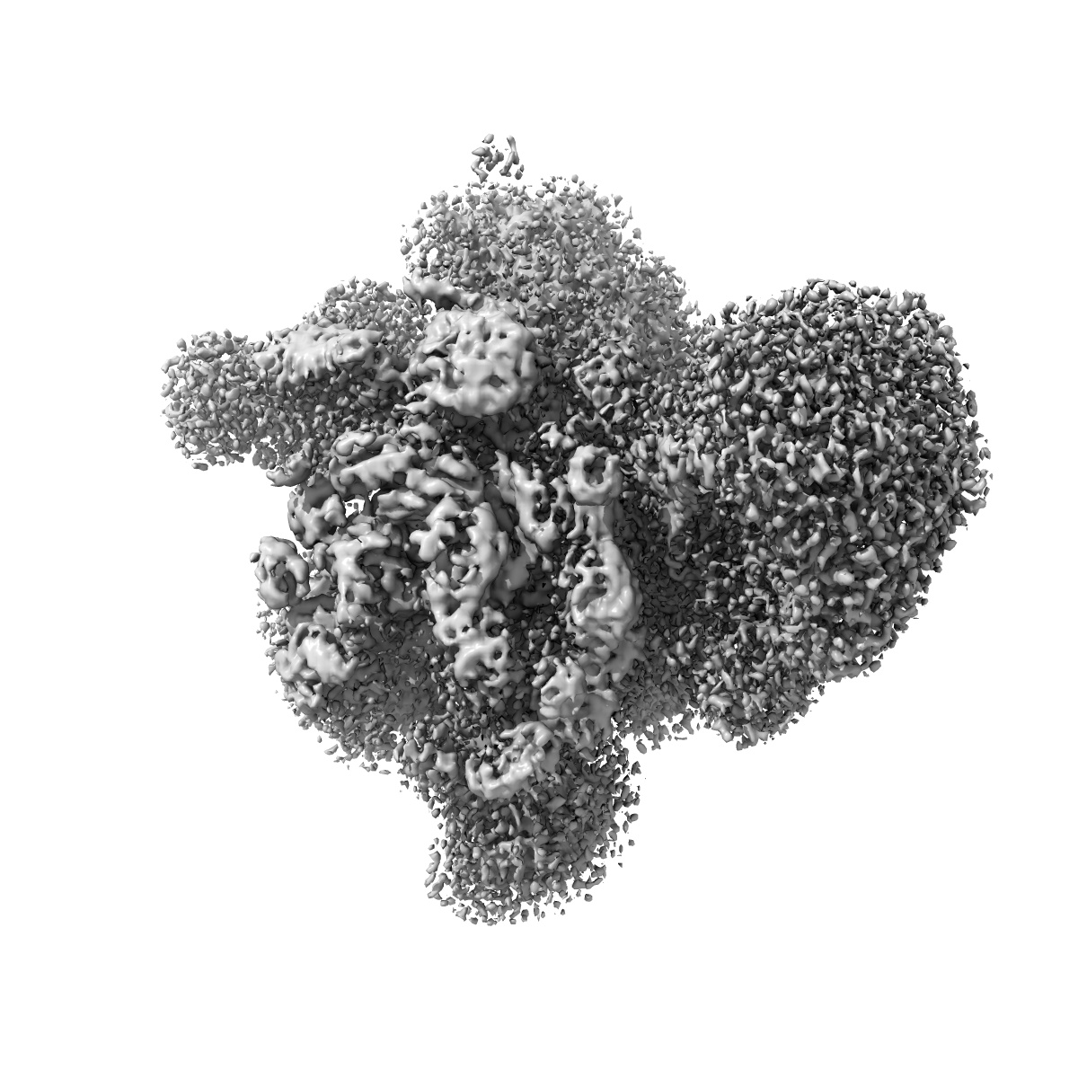
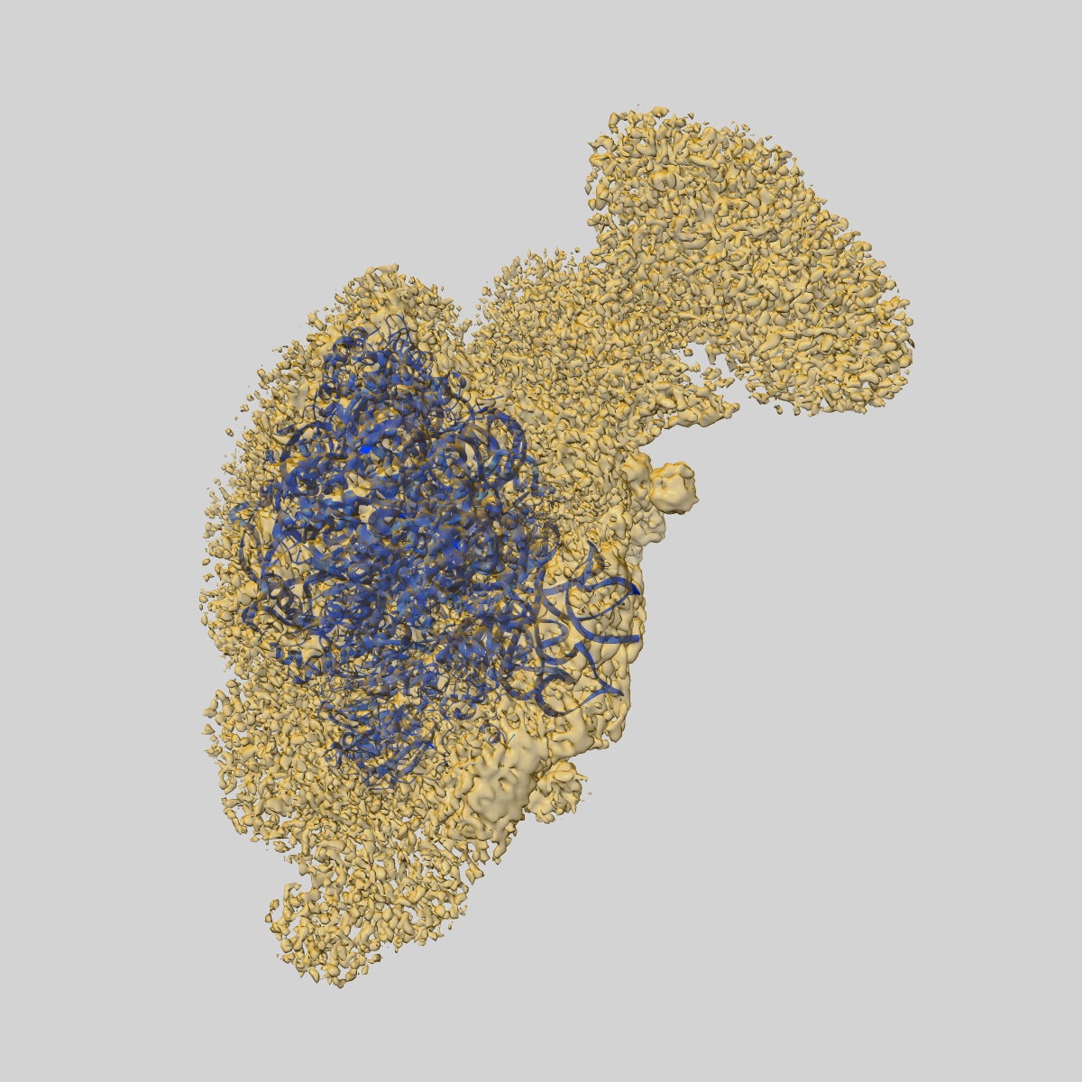
EMD-10772
Structure of a human 48S translational initiation complex - head
EMD-10772
Single-particle3.1 Å
 Deposition: 17/03/2020
Map released: 16/09/2020
Last modified: 24/04/2024
Buffer
pH: 7.4
Vitrification
Microscope: FEI TITAN KRIOS
Illumination mode: FLOOD BEAM
Imaging mode: BRIGHT FIELD
Electron source: FIELD EMISSION GUN
Acceleration voltage: 300 kV
Illumination mode: FLOOD BEAM
Imaging mode: BRIGHT FIELD
Electron source: FIELD EMISSION GUN
Acceleration voltage: 300 kV
Image Recording
[1]
Detector model:
FEI FALCON III (4k x 4k)
Detector mode: INTEGRATING
Average exposure time: 1.0 s
Average electron dose per image: 107.0 e/Å2
Detector mode: INTEGRATING
Average exposure time: 1.0 s
Average electron dose per image: 107.0 e/Å2
Format: CCP4
Data type: IMAGE STORED AS FLOATING POINT NUMBER (4 BYTES)
Data type: IMAGE STORED AS FLOATING POINT NUMBER (4 BYTES)
⬡ Geometry
| X | Y | Z | |
|---|---|---|---|
| Dimensions | 500 | 500 | 500 |
| Origin | 0 | 0 | 0 |
| Spacing | 500 | 500 | 500 |
| Voxel size | 1.074 Å | 1.074 Å | 1.074 Å |
Contour list
| Primary | Level | Source |
|---|---|---|
| True | 0.02 | AUTHOR |