EMD-10519
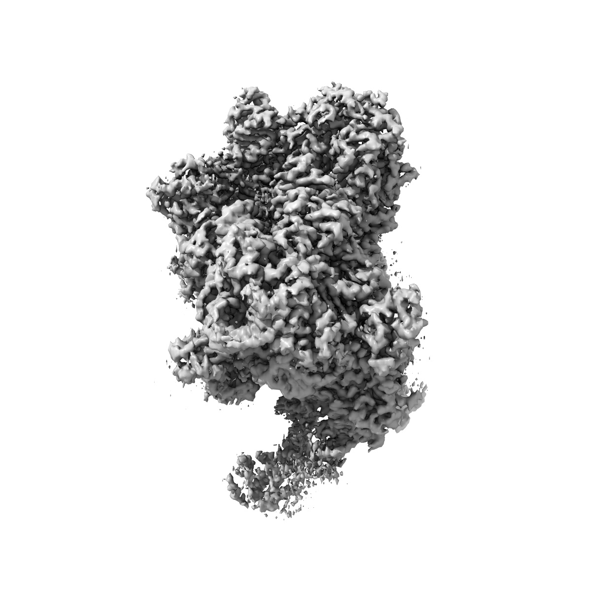
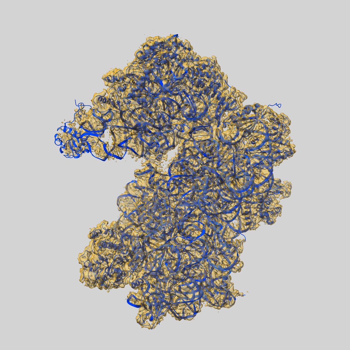
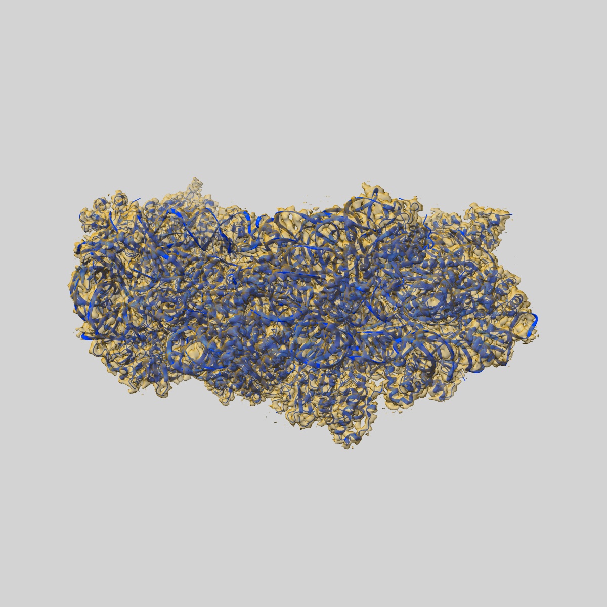
Structure of an archaeal ABCE1-bound ribosomal post-splitting complex
EMD-10519
Single-particle2.8 Å
 Deposition: 04/12/2019
Map released: 12/02/2020
Last modified: 02/12/2020
Buffer
pH: 7.5
Vitrification
Cryogen name: ETHANE
Microscope: FEI TITAN KRIOS
Illumination mode: FLOOD BEAM
Imaging mode: BRIGHT FIELD
Electron source: FIELD EMISSION GUN
Acceleration voltage: 300 kV
Illumination mode: FLOOD BEAM
Imaging mode: BRIGHT FIELD
Electron source: FIELD EMISSION GUN
Acceleration voltage: 300 kV
Image Recording
[1]
Final
reconstruction
Resolution: 2.8
Å
(
BY AUTHOR)
Resolution method: FSC 0.143 CUT-OFF
Number of images used: 293010
Resolution method: FSC 0.143 CUT-OFF
Number of images used: 293010
⌯ Applied Symmetry
Point group:
C1
Software
[1]
| Name | Version | Details |
|---|---|---|
| RELION | 3 | - |
⦨ Initial angle
assignment
Type:
MAXIMUM LIKELIHOOD
⦩ Final angle assignment
Type:
MAXIMUM LIKELIHOOD
Format: CCP4
Data type: IMAGE STORED AS FLOATING POINT NUMBER (4 BYTES)
Annotation details: Composite map of post-processed maps
Data type: IMAGE STORED AS FLOATING POINT NUMBER (4 BYTES)
Annotation details: Composite map of post-processed maps
⬡ Geometry
| X | Y | Z | |
|---|---|---|---|
| Dimensions | 360 | 360 | 360 |
| Origin | 0 | 0 | 0 |
| Spacing | 360 | 360 | 360 |
| Voxel size | 1.084 Å | 1.084 Å | 1.084 Å |
Contour list
| Primary | Level | Source |
|---|---|---|
| True | 5.0 | AUTHOR |