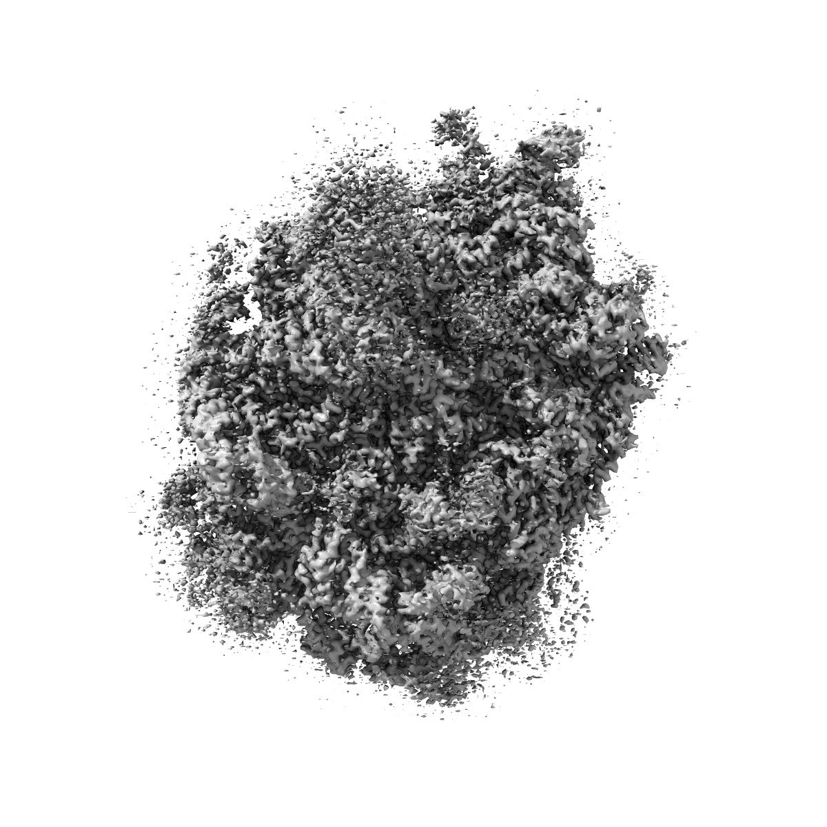
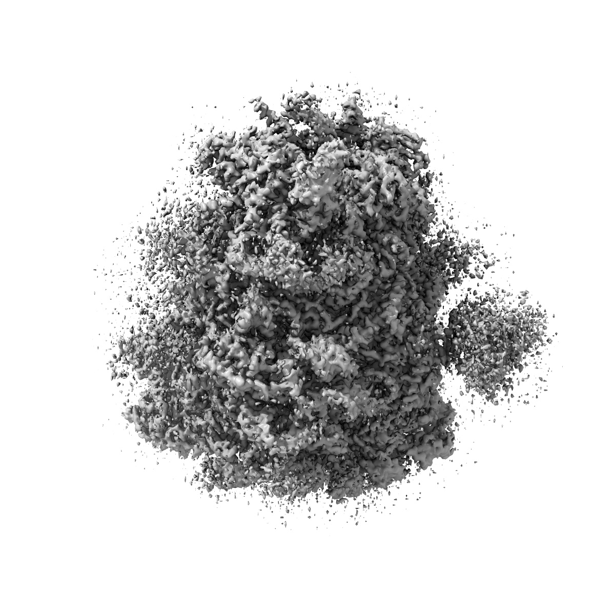
EMD-10380
Structure of rabbit 80S ribosome translating beta-tubulin in complex with tetratricopeptide protein 5 and nascent chain-associated complex
EMD-10380
Single-particle3.11 Å
 Deposition: 15/10/2019
Map released: 27/11/2019
Last modified: 02/12/2020
Buffer
pH: 7.4
Vitrification
Cryogen name: ETHANE
Microscope: FEI TITAN KRIOS
Illumination mode: FLOOD BEAM
Imaging mode: BRIGHT FIELD
Electron source: FIELD EMISSION GUN
Acceleration voltage: 300 kV
Illumination mode: FLOOD BEAM
Imaging mode: BRIGHT FIELD
Electron source: FIELD EMISSION GUN
Acceleration voltage: 300 kV
Image Recording
[1]
Detector model:
FEI FALCON III (4k x 4k)
Detector mode: INTEGRATING
Average electron dose per image: 48.36 e/Å2
Detector mode: INTEGRATING
Average electron dose per image: 48.36 e/Å2
Final
reconstruction
Resolution: 3.11
Å
(
BY AUTHOR)
Resolution method: FSC 0.143 CUT-OFF
Number of images used: 49626
Resolution method: FSC 0.143 CUT-OFF
Number of images used: 49626
⌯ Applied Symmetry
Point group:
C1
⦨ Initial angle
assignment
Type:
MAXIMUM LIKELIHOOD
⦩ Final angle assignment
Type:
MAXIMUM LIKELIHOOD
Format: CCP4
Data type: IMAGE STORED AS FLOATING POINT NUMBER (4 BYTES)
Annotation details: Structure of human TTC5 bound to the rabbit 80S ribosome translating beta-tubulin Sharpened, MTF-corrected map with the calibrated pixel size
Data type: IMAGE STORED AS FLOATING POINT NUMBER (4 BYTES)
Annotation details: Structure of human TTC5 bound to the rabbit 80S ribosome translating beta-tubulin Sharpened, MTF-corrected map with the calibrated pixel size
⬡ Geometry
| X | Y | Z | |
|---|---|---|---|
| Dimensions | 400 | 400 | 400 |
| Origin | 0 | 0 | 0 |
| Spacing | 400 | 400 | 400 |
| Voxel size | 1.33 Å | 1.33 Å | 1.33 Å |
Contour list
| Primary | Level | Source |
|---|---|---|
| True | 0.08 | AUTHOR |