EMD-0987
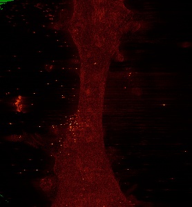
Ultra-high voltage electron microscope tomography using 1000-nm-thick neurite section acquired at 20,000 magnification at an accelerating voltage of 2 MV
EMD-0987
Tomography Deposition: 01/02/2020
Map released: 20/05/2020
Last modified: 20/05/2020
Buffer
pH: 7.3
Staining
Sugar
embedding
Material: Resin
Fiducial Markers
| Manufacturer | Fiducial type | Diameter |
|---|---|---|
| BBI Solutions | - | 20 nanometer |
Sectioning (Ultramicrotomy)
Microscope: HITACHI H3000 UHVEM
Illumination mode: FLOOD BEAM
Imaging mode: BRIGHT FIELD
Electron source: LAB6
Acceleration voltage: 2000 kV
Nominal magnification: 20000.0
Specimen holder model: OTHER
Illumination mode: FLOOD BEAM
Imaging mode: BRIGHT FIELD
Electron source: LAB6
Acceleration voltage: 2000 kV
Nominal magnification: 20000.0
Specimen holder model: OTHER
Image Recording
[1]
Final
reconstruction
Format: CCP4
Data type: IMAGE STORED AS SIGNED INTEGER (2 BYTES)
Annotation details: 1.0-um-thick neurite section of cultured PC12 cells(Magnification 20K)
Data type: IMAGE STORED AS SIGNED INTEGER (2 BYTES)
Annotation details: 1.0-um-thick neurite section of cultured PC12 cells(Magnification 20K)
⬡ Geometry
| X | Y | Z | |
|---|---|---|---|
| Dimensions | 934 | 1008 | 91 |
| Origin | -2 | 3 | 2 |
| Spacing | 934 | 1008 | 91 |
| Voxel size | 34.0 Å | 34.0 Å | 34.0 Å |
Contour list
| Primary | Level | Source |
|---|---|---|
| True | - | AUTHOR |