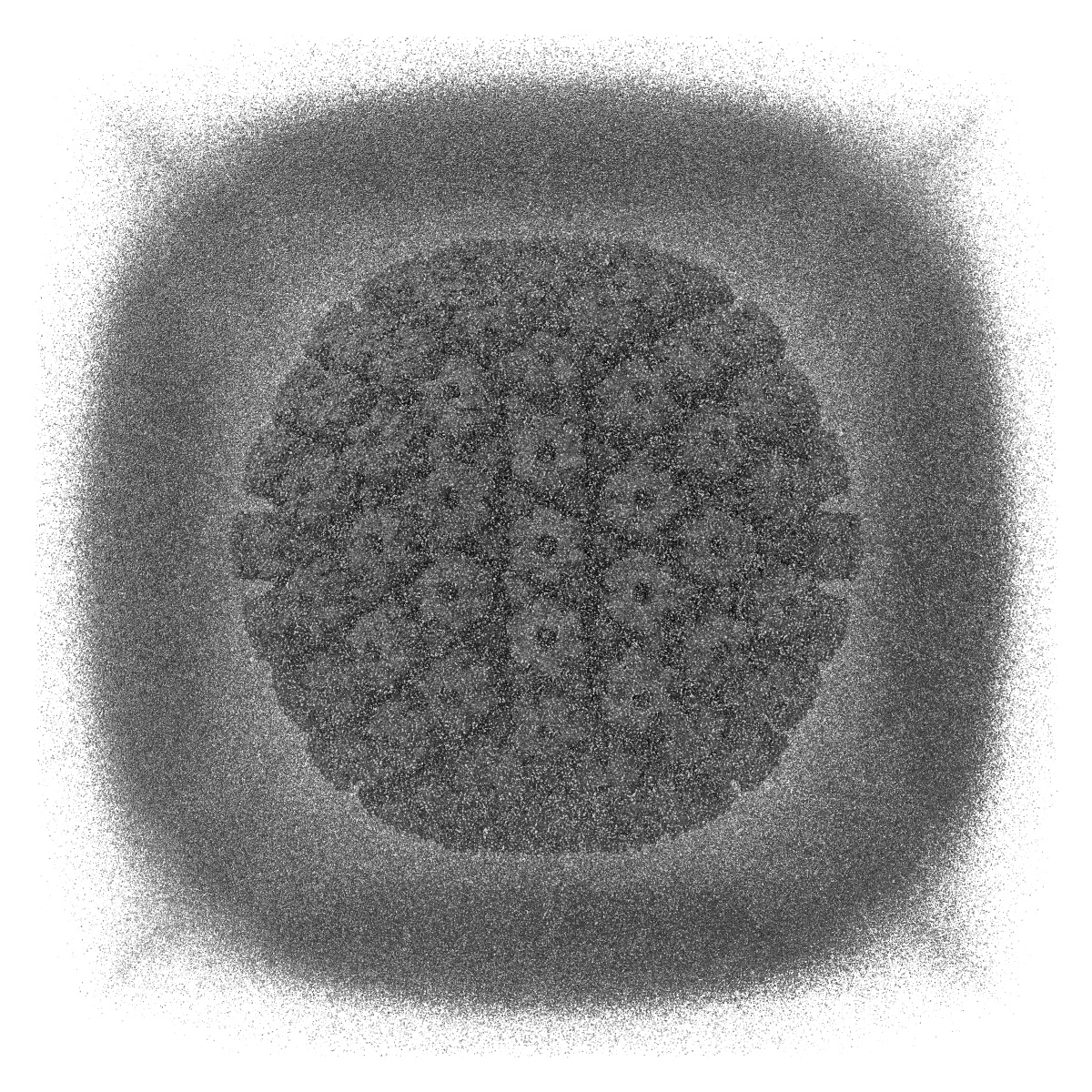
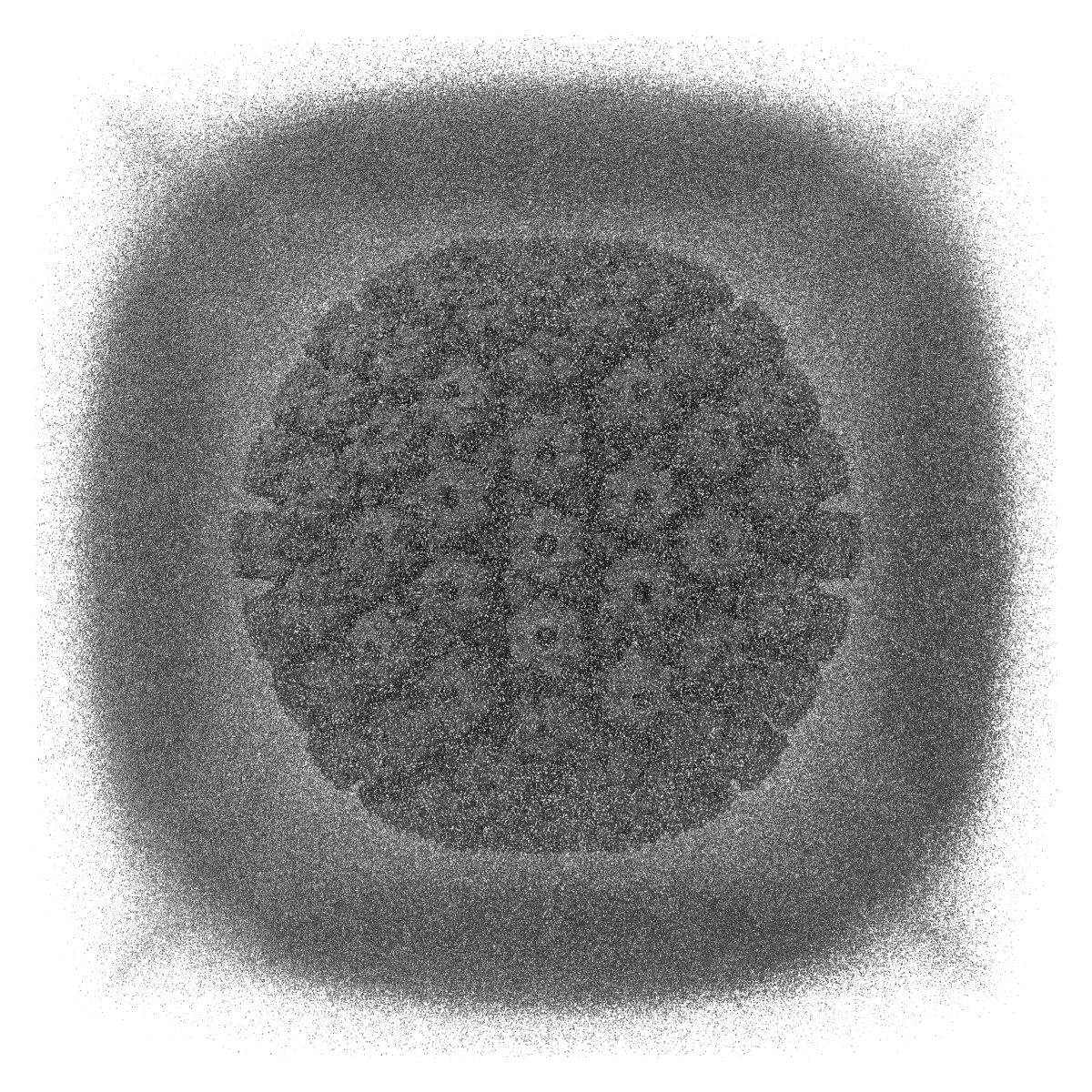
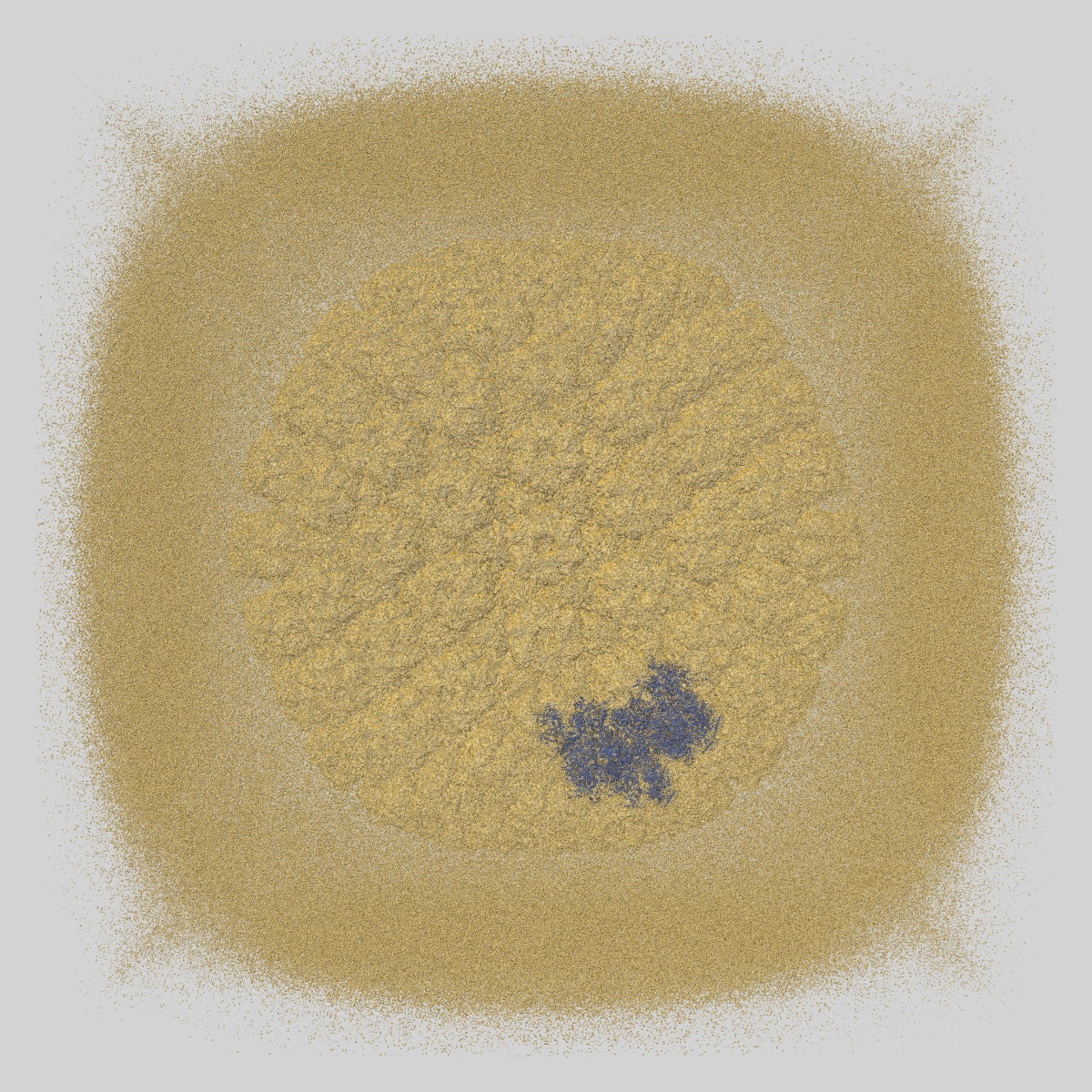
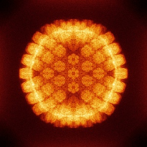
EMD-0880
The atomic structure of varicella-zoster virus A-capsid
EMD-0880
Single-particle4.4 Å
 Deposition: 05/12/2019
Map released: 29/07/2020
Last modified: 11/08/2021
Buffer
pH: 7.4
Vitrification
Cryogen name: ETHANE
Microscope: FEI TITAN KRIOS
Illumination mode: FLOOD BEAM
Imaging mode: BRIGHT FIELD
Electron source: FIELD EMISSION GUN
Acceleration voltage: 300 kV
Illumination mode: FLOOD BEAM
Imaging mode: BRIGHT FIELD
Electron source: FIELD EMISSION GUN
Acceleration voltage: 300 kV
Image Recording
[1]
Detector model:
GATAN K2 SUMMIT (4k x 4k)
Detector mode: SUPER-RESOLUTION
Average electron dose per image: 56.0 e/Å2
Detector mode: SUPER-RESOLUTION
Average electron dose per image: 56.0 e/Å2
Final
reconstruction
Resolution: 4.4
Å
(
BY AUTHOR)
Resolution method: FSC 0.143 CUT-OFF
Number of images used: 22983
Resolution method: FSC 0.143 CUT-OFF
Number of images used: 22983
⌯ Applied Symmetry
Point group:
I
⦨ Initial angle
assignment
Type:
PROJECTION MATCHING
⦩ Final angle assignment
Type:
MAXIMUM LIKELIHOOD
Format: CCP4
Data type: IMAGE STORED AS FLOATING POINT NUMBER (4 BYTES)
Annotation details: Cryo-EM structure of varicella-zoster virus A-capsid
Data type: IMAGE STORED AS FLOATING POINT NUMBER (4 BYTES)
Annotation details: Cryo-EM structure of varicella-zoster virus A-capsid
⬡ Geometry
| X | Y | Z | |
|---|---|---|---|
| Dimensions | 1280 | 1280 | 1280 |
| Origin | 0 | 0 | 0 |
| Spacing | 1280 | 1280 | 1280 |
| Voxel size | 1.307 Å | 1.307 Å | 1.307 Å |
Contour list
| Primary | Level | Source |
|---|---|---|
| True | 3.0 | AUTHOR |