The image for April in our 2018 calendar depicts a single Zika virus protein in addition to the full Zika virus capsid. In this feature we discuss the transition between these two structures and demonstrate the beautiful simplicity of virus assembly.
The assembly of virus particles is a mammoth task, involving the complex organisation of anywhere from 60 to 10,000 protein subunits into a single structure called the viral capsid. This encloses the viral genome, protecting it and transporting it to infect new host cells. The capsid can vary greatly in size and complexity, with recently discovered giant viruses (e.g. Faustovirus) being larger than some bacteria! The key to building these giant structures is in their simplicity. The viral capsid is formed from only a small number of different proteins, which in turn form only a limited number of arrangements, repeating to create the overall capsid.
Shaping the virus capsid
The differences in the way individual protein subunits assemble, affecting capsid size and shape, can be easily demonstrated using a few examples. Firstly, and most basically, icosahedral viruses classified as T=1, which have subunits in only one unique environment. These capsids contain a single type of protein that assembles into triangles (see figure 1, left). This basic shape is then repeated to generate the 5-fold symmetry of the icosahedron from which the type of virus gets its name. This is the simplest arrangement in an icosahedral virus and generally produces the smallest virus capsids, around 20 nm in diameter. The example shown is that of Satellite Tobacco mosaic virus (PDB entry 2buk).

Figure 1: Left, structure of T-1 class virus with triangular and pentameric geometry shown. Right, structure of T-7 class virus with triangular, pentameric and hexagonal geometry shown. Inset, at center top, shows a football built from pentameric and hexagonal geometry.
Alternative arrangement of subunits in icosahedral viruses can enable much larger capsid formation, yet are only slightly more complex, maintaining a basic subunit structure with 60 repeating copies (figure above) which can be thought of as assembly of pentamers and hexagons, much like a football. Viruses classified as T=7 still arrange their capsid proteins into triangles, however there are seven unique subunit environments, giving an overall assembly of 420 subunits. The subunits are again arranged into a pentameric structure, as in T=1 capsids, however each is surrounded by an arrangement of hexagons formed from the additional subunits in the basic triangular unit. This, in effect, spreads out the pentameric apices of the icosahedron, leading to larger faces on the virus and consequently a larger viral capsid structure. Examples of these arrangements of viral subunits includes the head unit of many bacteriophages, including the Bacteriophage HK97 (PDB entry 1ohg), which is shown in figure 1, right. The difference in size of these two viruses can be seen in figure 2, indicating how this subtle change in subunit organisation can significantly alter the proportions of the capsid.

Figure 2: Size comparison between a T=1 viral capsid structure, Satellite Tobacco mosaic virus, and a T=7 viral capsid, Bacteriophage HK97.
These different subunit arrangements can also enable more elaborate viral capsid structures to form from a limited number of subunits. For example, the HIV capsid has an irregular cone-like shape, as can be seen in figure 3. The underlying reason for this capsid structure can be seen by the arrangement of subunits into either hexagons or pentagons. The more gently curved regions of the capsid contain only the hexagonal structures, whereas occasional pentameric arrangements can be seen at more highly curved regions of the capsid. This irregular combination of different structures produces a unique capsid structure that may be important for the virus in preventing immune attack.

Figure 3: Structure of HIV capsid from side view (left) and end view (right), with the different geometrtic shapes displayed in black dashed lines.
Zika structure
The Zika virus is classified as T=3, similar to that of Dengue virus. Its capsid forms from a repeating structure of 3 protein chains, forming pentagonal shapes surrounded by hexagons. Each hexagon in this case is in contact with three pentagonal apices, thus forming a more expanded structure than T=1 viruses but more compact than the T=7 viruses. The 3D structure of the virus has been determined in near-atomic detail by cryo-electron microscopy (PDB entry 5ire). The structure indicates that, while similar to Dengue virus, Zika differs in the regions where carbohydrates are attached to the virus surface. These regions and the carbohydrates might affect how the virus infects human cells, and the authors of the paper describing the structure hypothesise that the carbohydrates may function as attachment sites of the virus to host cells. For information about Zika virus and its structure, see our previous featured structure.
Zika activities
You can make your own Zika virus, using our virus net template. Just cut out the net, fold along the lines and stick the virus together using the tabs.
You can also view the virus in augmented reality, using the Augment app on your tablet or smartphone. If you have a copy of our 2018 calendar then just scan the page for April and the structure will appear above the page! You can also view the augmented reality structure directly by following this link.
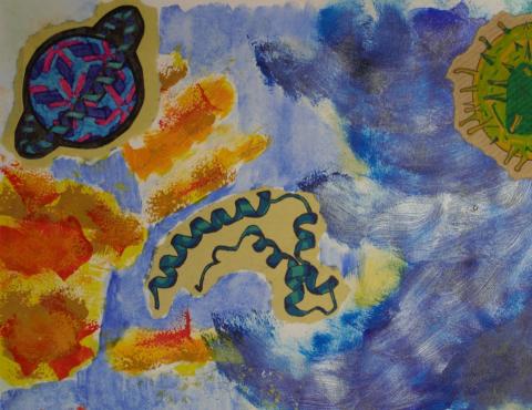
Artwork
The image in our calendar for April shows the Zika virus and components presented in an abstract environment. The artwork was created by student Emily Harris from Impington Village College, Cambridge using acrylics and ink. Here the story is told in the form of a poem, by the artist from the perspective of the virus:
“Drifting, floating innocently, in the depth of a crimson sea.
I know I am destined to kill, but still I have hopeless good will.
Stay away if you want to survive, or deep within your veins I will thrive.
Hidden, unseen, cursed hands unclean, I hide in plain sight, destination bloodstream.”