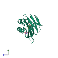
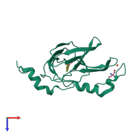
6vi7

X-ray diffraction
2.62Å resolution

Probing extradiol dioxygenase mechanism in NAD(+) biosynthesis by viewing reaction cycle intermediates - a substrate bidentately bound structure

Released:
Primary publication:
Observing 3-hydroxyanthranilate-3,4-dioxygenase in action through a crystalline lens.
Proc Natl Acad Sci U S A 117 19720-19730 (2020)
PMID: 32732435

Function and Biology Details

Reaction catalysed:
3-hydroxyanthranilate + O(2) = 2-amino-3-carboxymuconate semialdehyde
Biochemical function:
Biological process:
Cellular component:

Structure analysis Details

Assembly composition:
homo dimer (preferred)
PDBe Complex ID:
PDB-CPX-172948 (preferred)
Entry contents:
1 distinct polypeptide molecule
Macromolecule:
3-hydroxyanthranilate 3,4-dioxygenase Chain: A
Molecule details ›
Chain: A
Length: 195 amino acids
Theoretical weight: 22.59 KDa
Source organism: Cupriavidus metallidurans CH34
Expression system: Escherichia coli BL21(DE3)
UniProt:
  • Canonical: Q1LCS4 (Residues: 1-174; Coverage: 100%)
Gene names: Rmet_5193, nbaC
Sequence domains: 3-hydroxyanthranilic acid dioxygenase

Ligands and Environments

3 bound ligands:
No modified residues

Experiments and Validation Details

Entry percentile scores
X-ray source: APS BEAMLINE 19-BM
Spacegroup: P6522
Unit cell:
a: 58.04Å b: 58.04Å c: 231.58Å
α: 90° β: 90° γ: 120°
R-values:
R R work R free
0.208 0.199 0.29
Expression system: Escherichia coli BL21(DE3)