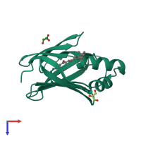
6mpk

X-ray diffraction
1.58Å resolution

Crystal Structure of the 13-cis Retinal-Bound R111K:Y134F:T54V:R132Q:P39Y:R59Y:L121E:A32Y Mutant of Human Cellular Retinoic Acid Binding Protein II in the Dark at 1.58 Angstrom Resolution

Released:
Source organism: Homo sapiens

Function and Biology Details

Structure analysis Details

Assembly composition:
monomeric (preferred)
PDBe Complex ID:
PDB-CPX-151430 (preferred)
Entry contents:
1 distinct polypeptide molecule
Macromolecule:
Cellular retinoic acid-binding protein 2 Chain: A
Molecule details ›
Chain: A
Length: 137 amino acids
Theoretical weight: 15.69 KDa
Source organism: Homo sapiens
Expression system: Bacterial expression vector pBEN1-SGC
UniProt:
  • Canonical: P29373 (Residues: 2-138; Coverage: 99%)
Gene name: CRABP2
Sequence domains: Lipocalin / cytosolic fatty-acid binding protein family
Structure domains: Lipocalin

Ligands and Environments

2 bound ligands:
No modified residues

Experiments and Validation Details

Entry percentile scores
X-ray source: APS BEAMLINE 21-ID-F
Spacegroup: P3121
Unit cell:
a: 58.761Å b: 58.761Å c: 99.174Å
α: 90° β: 90° γ: 120°
R-values:
R R work R free
0.204 0.203 0.222
Expression system: Bacterial expression vector pBEN1-SGC