
phenyl]biphenyl-4,4'-dicarboxamide</span>;</li> <li class='image_legend_li'>1 copy of <span class='highlight'>PHOSPHATE ION</span>;</li> <li class='image_legend_li'>1 copy of <span class='highlight'>water</span>.</li></ul>")
phenyl]biphenyl-4,4'-dicarboxamide</span>;</li> <li class='image_legend_li'>1 copy of <span class='highlight'>PHOSPHATE ION</span>;</li> <li class='image_legend_li'>1 copy of <span class='highlight'>water</span>.</li></ul>")
phenyl]biphenyl-4,4'-dicarboxamide</span>;</li> <li class='image_legend_li'>1 copy of <span class='highlight'>PHOSPHATE ION</span>;</li> <li class='image_legend_li'>1 copy of <span class='highlight'>water</span>.</li></ul>")
Function and Biology Details
Reactions catalysed:
Prenyl diphosphate + isopentenyl diphosphate = diphosphate + geranyl diphosphate
Geranyl diphosphate + isopentenyl diphosphate = diphosphate + (2E,6E)-farnesyl diphosphate
Biochemical function:
Biological process:
Cellular component:
- not assigned
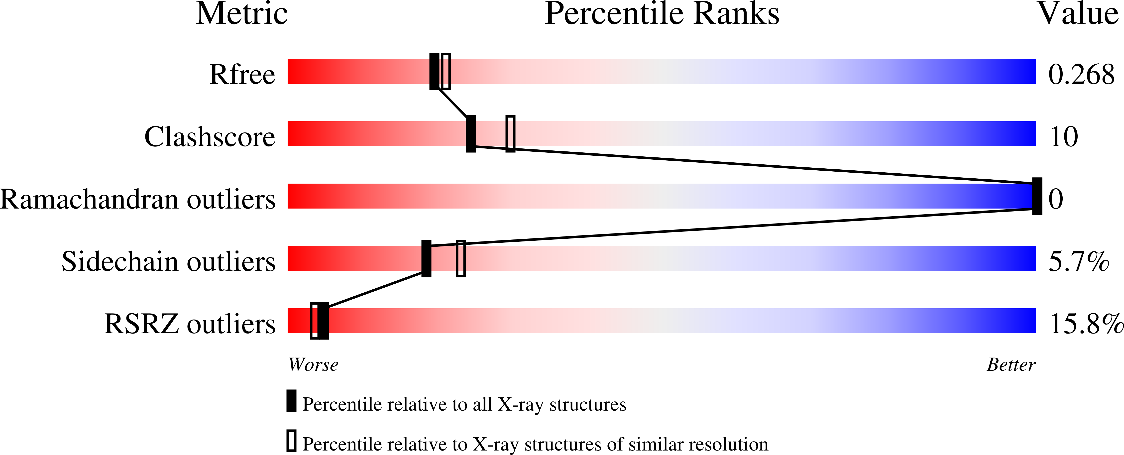
Structure analysis Details
Assembly composition:
homo dimer (preferred)
Assembly name:
Farnesyl pyrophosphate synthase (preferred)
PDBe Complex ID:
PDB-CPX-146902 (preferred)
Entry contents:
1 distinct polypeptide molecule
Macromolecule:
Farnesyl pyrophosphate synthase
Molecule details ›
Chain: A
Length: 348 amino acids
Theoretical weight: 40.03 KDa
Source organism: Homo sapiens
Expression system: Escherichia coli
UniProt:
Sequence domains: Polyprenyl synthetase
Structure domains: Farnesyl Diphosphate Synthase
Length: 348 amino acids
Theoretical weight: 40.03 KDa
Source organism: Homo sapiens
Expression system: Escherichia coli
UniProt:
- Canonical:
 P14324 (Residues: 72-419; Coverage: 83%)
Sequence domains: Polyprenyl synthetase
Structure domains: Farnesyl Diphosphate Synthase


