


Function and Biology Details
Biochemical function:
- not assigned
Biological process:
- not assigned
Cellular component:
- not assigned
Structure analysis Details
Assembly composition:
hetero dimer (preferred)
Assembly name:
PDBe Complex ID:
PDB-CPX-152649 (preferred)
Entry contents:
2 distinct polypeptide molecules
Macromolecules (2 distinct):
Coatomer subunit beta'
Molecule details ›
Chains: A, B
Length: 301 amino acids
Theoretical weight: 34.29 KDa
Source organism: Saccharomyces cerevisiae
UniProt:
Sequence domains: WD domain, G-beta repeat
Structure domains: YVTN repeat-like/Quinoprotein amine dehydrogenase
Length: 301 amino acids
Theoretical weight: 34.29 KDa
Source organism: Saccharomyces cerevisiae
UniProt:
- Canonical:
 P41811 (Residues: 1-301; Coverage: 34%)
Sequence domains: WD domain, G-beta repeat
Structure domains: YVTN repeat-like/Quinoprotein amine dehydrogenase
Dolichyl-diphosphooligosaccharide--protein glycosyltransferase subunit WBP1
Molecule details ›
Chains: C, D
Length: 6 amino acids
Theoretical weight: 740 Da
Source organism: Saccharomyces cerevisiae
Expression system: Not provided
UniProt:
Length: 6 amino acids
Theoretical weight: 740 Da
Source organism: Saccharomyces cerevisiae
Expression system: Not provided
UniProt:
- Canonical: P33767 (Residues: 425-430; Coverage: 2%)
Ligands and Environments
No bound ligands
No modified residues
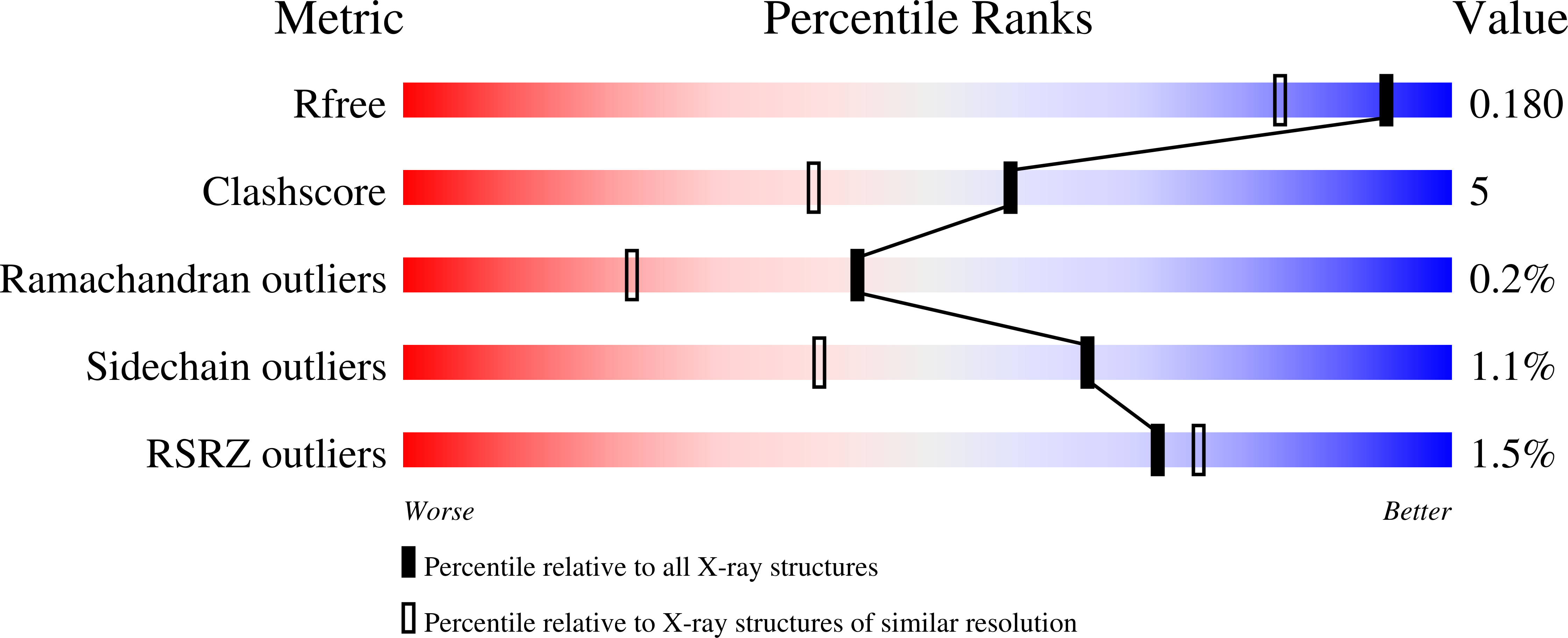
Experiments and Validation Details

X-ray source:
APS BEAMLINE 24-ID-E
Spacegroup:
P21
Expression system: Not provided