Function and Biology Details
Reaction catalysed:
An aliphatic amide = a nitrile + H(2)O
Biochemical function:
Biological process:
Cellular component:
- not assigned
Sequence domains:
- Nitrile hydratase alpha /Thiocyanate hydrolase gamma superfamily
- Nitrile hydratase beta subunit, N-terminal
- Nitrile hydratase beta subunit domain
- Electron transport accessory-like domain superfamily
- Nitrile hydratase, beta subunit
- Nitrile hydratase alpha /Thiocyanate hydrolase gamma
- Nitrile hydratase, alpha subunit
- Nitrile hydratase alpha subunit /Thiocyanate hydrolase gamma subunit
Structure analysis Details
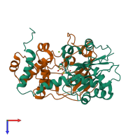
Assembly composition:
hetero dimer (preferred)
Assembly name:
Nitrile hydratase (preferred)
PDBe Complex ID:
PDB-CPX-146592 (preferred)
Entry contents:
2 distinct polypeptide molecules
Macromolecules (2 distinct):


