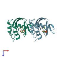
3jw1

X-ray diffraction
1.6Å resolution

Crystal Structure of Bovine Pancreatic Ribonuclease Complexed with Uridine-5'-monophosphate at 1.60 A Resolution

Released:
Source organism: Bos taurus
Primary publication:
Structure of bovine pancreatic ribonuclease complexed with uridine 5'-monophosphate at 1.60 A resolution.
Acta Crystallogr Sect F Struct Biol Cryst Commun 66 113-20 (2010)
PMID: 20124705

Function and Biology Details

Reaction catalysed:
(1a) an (RNA) containing cytidine = an (RNA)-3'-cytidine-2',3'-cyclophosphate + a 5'-hydroxy-ribonucleotide-3'-(RNA)
Biochemical function:
Cellular component:

Structure analysis Details

Assembly composition:
monomeric (preferred)
PDBe Complex ID:
PDB-CPX-158390 (preferred)
Entry contents:
1 distinct polypeptide molecule
Macromolecule:
Ribonuclease pancreatic Chains: A, B
Molecule details ›
Chains: A, B
Length: 124 amino acids
Theoretical weight: 13.71 KDa
Source organism: Bos taurus
UniProt:
  • Canonical: P61823 (Residues: 27-150; Coverage: 100%)
Gene names: RNASE1, RNS1
Sequence domains: Pancreatic ribonuclease
Structure domains: Ribonuclease A-like domain

Ligands and Environments

1 bound ligand:
No modified residues

Experiments and Validation Details

Entry percentile scores
X-ray source: RIGAKU RU200
Spacegroup: P21
Unit cell:
a: 30.73Å b: 74.85Å c: 50.52Å
α: 90° β: 107.8° γ: 90°
R-values:
R R work R free
0.203 0.197 0.253