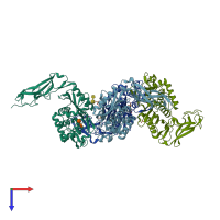
Function and Biology Details
Reaction catalysed:
Preferential cleavage: (Ac)(2)-L-Lys-D-Ala-|-D-Ala. Also transpeptidation of peptidyl-alanyl moieties that are N-acyl substituents of D-alanine.
Biochemical function:
Biological process:
Cellular component:
Sequence domains:
- Peptidase S11, D-alanyl-D-alanine carboxypeptidase A
- Beta-lactamase/transpeptidase-like
- D-Ala-D-Ala carboxypeptidase, C-terminal domain superfamily
- Penicillin-binding protein, C-terminal domain superfamily
- Peptidase S11, D-Ala-D-Ala carboxypeptidase A, C-terminal
- Peptidase S11, D-alanyl-D-alanine carboxypeptidase A, N-terminal
Structure analysis Details
Assembly composition:
monomeric (preferred)
Assembly name:
D-alanyl-D-alanine carboxypeptidase DacC (preferred)
PDBe Complex ID:
PDB-CPX-140150 (preferred)
Entry contents:
1 distinct polypeptide molecule
Macromolecules (2 distinct):


